Download
1 / 18
190 likes | 670 Vues
Measurement of Haemoglobin. Dr Maliha Sumbul. Hemoglobin ( also spelled haemoglobin and abbreviated Hb or Hgb ) is the iron -containing oxygen -transport metalloprotein in the red blood cells of vertebrates , [1] and the tissues of some invertebrates .

E N D
Measurement of Haemoglobin Dr Maliha Sumbul
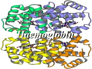
Hemoglobin (also spelledhaemoglobin and abbreviated Hb or Hgb) is the iron-containing oxygen-transport metalloprotein in the red blood cells of vertebrates,[1] and the tissues of some invertebrates. • In mammals, the protein makes up about 97% of the red blood cell’s dry content, and around 35% of the total content (including water). Hemoglobin transports oxygen from the lungs or gills to the rest of the body where it releases the oxygen for cell use. It also has a variety of other roles of gas transport and effect-modulation which vary from species to species, and are quite diverse in some invertebrates. • Hemoglobin has an oxygen binding capacity of between 1.36 and 1.37 ml O2 per gram hemoglobin,[2] which increases the total blood oxygen capacity seventyfold.[3] • Hemoglobin is found in nonerythroid cells including the A9 dopaminergic neurons in the substantia nigra, macrophages, alveolar cells, and mesangial cells in the kidney. In these tissues, it has a non-oxygen carrying function as an antioxidant and a regulator of iron metabolism.[4]
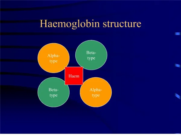
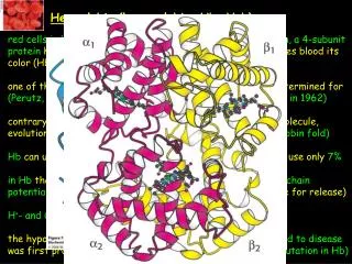
Structure • Heme group • In most humans, the hemoglobin molecule is an assembly of four globular protein subunits. Each subunit is composed of a protein chain tightly associated with a non-protein heme group. Each protein chain arranges into a set of alpha-helix structural segments connected together in a globin fold arrangement, so called because this arrangement is the same folding motif used in other heme/globin proteins such as myoglobin.[15][16] This folding pattern contains a pocket which strongly binds the heme group. • A heme group consists of an iron (Fe) ion (charged atom) held in a heterocyclic ring, known as a porphyrin. The iron ion, which is the site of oxygen binding, coordinates with the four nitrogens in the center of the ring, which all lie in one plane. The iron is also bound strongly to the globular protein via the imidazole ring of the F8 histidine residue below the porphyrin ring. A sixth position can reversibly bind oxygen by a coordinate covalent bond[17], completing the octahedral group of six ligands. Oxygen binds in an "end-on bent" geometry where one oxygen atom binds Fe and the other protrudes at an angle. When oxygen is not bound, a very weakly bonded water molecule fills the site, forming a distorted octahedron. • The iron ion may either be in the Fe2+ or Fe3+ state, but ferrihemoglobin (methemoglobin) (Fe3+) cannot bind oxygen.[18] In binding, oxygen temporarily oxidizes (Fe2+) to (Fe3+), so iron must exist in the +2 oxidation state in order to bind oxygen.
A schematic visual model of oxygen binding process, showing all four monomers and hemes , and protein chains only as diagramatic coils, to facilitate visualization into the molecule. Oxygen is not shown in this model, but for each of the iron atoms it binds to the iron (red sphere) in the flat heme. For example, in the upper left of the four hemes shown, oxygen binds at the left of the iron atom shown in the upper left of diagram. This causes the iron atom to move backward into the heme which holds it, tuging the histidine residue (modeled as a red pentagon on the right of the iron) closer, as it does. This, in turn, pulls on the protein chain holding the histidine.
Heart of Steel (Hemoglobin) (2005) by Julian Voss-Andreae. The images show the 5' (1.60 m) tall sculpture right after installation, after 10 days, and after several months of exposure to the elements.
In history , art and music • Historically, the color of blood was associated with rust, as ancient Romans associated the planet Mars with the god of war since Mars is orange-red. The color of Mars is due to iron-oxygen in the Martian soil, but the red in blood is not due to the iron in hemoglobin and its oxides, which is a common misconception. The red is due to the porphyrinmoiety of hemoglobin to which the iron is bound, not the iron itself,[45] although the ligation and redox state of the iron can influence the pi to pi* electronic transitions of the porphyrin and hence its optical characteristics. • Heart of Steel (Hemoglobin) (2005) by Julian Voss-Andreae. The images show the 5' (1.60 m) tall sculpture right after installation, after 10 days, and after several months of exposure to the elements. • Artist Julian Voss-Andreae created a sculpture called "Heart of Steel (Hemoglobin)" in 2005, based on the protein's backbone. The sculpture was made from glass and weathering steel. The intentional rusting of the initially shiny work of art mirrors hemoglobin's fundamental chemical reaction of oxygen binding to iron.[46] • Rock band Placebo recorded a song called Haemoglobin with the lyrics "Haemoglobin is the key to a healthy heartbeat".
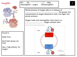
Principle of the test • WB + HiCN reagent ( Drabkin’s ) • K ferrricyanide converts Hb Fe from Fe++ Fe +++ forming met Hb (Hi) • Hi (metHb) combines with K cyanide to form stable pigment cyanmet Hb (HiCN) • Non – ionic detergent improves lysis of red cells and decreases turbidity due to abnormal proteins eg: lippproteins • Color intensity measured ( Spectrophotometer: 540 nm, Colorimeter: yellow green filter) O.D ( A) DIRECTLY PROPORTIONAL Hb Concentration oxidation
Calibration Curve • Prepared to check linearity of response of the instrument • Set up a series of 5 tubes • Into the tubes pipette sequentially 6.0, 4.5, 3.0, 1.5 and 0 ml of HiCN Reference Standard • Make the volumes up to 6 ml in each tube by adding, in the sequence, 0, 1.5, 3.0, 4.5 and 6 ml of reagent, giving, respectively, 100%, 75%, 50% and 25% of the original concentration of the reference standard
0% 25% 50% 75% 100% *Eg: 4.5/6.0 x 100
Measure the absorbance at 540 nm of each solution • Plot on arithmatic graph paper with Hb Concentration on the x – axis and absorbance on the y- axis • All points should fall on a line passing through zero • If there is irregularity in the graph, check the function of spectrophotometer
This check should be carried out whenever: - the spectrophotometer is serviced or repaired, - And also routinely approx. every 6 months
0.50 0.40 A 0.30 0.20 0.10 0 5 10 15 20 Hb conc
Calibration Table (Hb) • When many blood samples are tested each day, it is convenient to rwad results from a calibration graph ( or a table derived from it) that converts the readings of absorbance to Hb concentration as described above • In practice, readings will be reliable only if they lie within the linear part of the curve
continued • With most instruments, the most accurate and precise readings are in the range 0.2 – 0.7 absorbance units • If a reading occurs outside this range, the analysis should be repeated using a different, more appropriate, dilution of the sample • The calibration table is valid only for the instrument on which the values have been obtained • Each day, the table should be rechecked by measuring a sample from previous batch as well as a control preparation