Human Inheritance
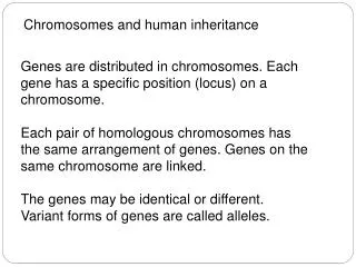
Human Inheritance. RECALL:. How many chromosomes do human body cells contain?. 46. YES!. Are there different types of chromosomes?. There are 2 types you should know: (WRITE THIS DOWN!). TYPES OF CHROMOSOMES Autosomes : (aka autosomal chromosomes) 22 homologous pairs in human cells.

Human Inheritance
E N D
Presentation Transcript
Human Inheritance RECALL: How many chromosomes do human body cells contain? 46 YES! Are there different types of chromosomes? There are 2 types you should know: (WRITE THIS DOWN!) • TYPES OF CHROMOSOMES • Autosomes: (aka autosomal chromosomes) 22 homologous pairs in human cells. • Sex chromosomes: X or Y, determine gender & contain genes important for male/female development.
Human Karyotypes • Biologists analyze the chromosomes of humans by trapping the cells in metaphase, the phase in which all 46 chromosomes can be seen, photographing them, cutting out the photograph of each chromosome and then grouping them together- this is known as a karyotype
Human Karyotypes • The karyotype reveals that each cell has 22 pairs of homologous chromosomes that are numbered from 1-22 in order of decreasing size • These 22 pairsare called autosomes, or autosomal chromosomes
SO...HOW DO YOU MAKE ONE? A karyotype of human chromosomes. Is this individual male or female?
How a human karyotype is made: • Cells are obtained from a fetus either by a) amniocentesis - removal of amniotic fluid surrounding the developing fetus b) chorionic villus sampling - tissue surrounding the fetus is removed and examined 2) The cell is “frozen” during mitosis with special chemicals. 3) Then the cell & its contents are treated with dyes (Giemsa stain) that will stain the chromosomes. 4) The cell is then burst open (spread) and the chromosomes are released and a picture is taken of the chromosomes. 5) Geneticists cut out the chromosomes from the enlarged picture & organize them by size, centromere location, & stain patterns.
Original Chromosome Smear: Cell was crushed/spread to release stained chromosomes.
Normal Female SEX CHROMOSOMES
Karyotypes are used to: • Determine if a developing baby may have genetic diseases or disorders due to abnormalities in chromosome number or shape. 2) Determine the gender of the baby. 3) Explain why a fetus was spontaneously aborted or died before birth. *They CANNOT show whether a baby is recessive or dominant for a particular trait-ONLY ANALYSIS OF DNA CAN! . LET’S TAKE A LOOK AT SOME OF THESE DISORDERS... SEE IF YOU CAN FIGURE OUT WHAT’S WRONG.
Normal Female SEX CHROMOSOMES
WHAT’S WRONG WITH THIS KARYOTYPE? IS SOMETHING MISSING?
Turner Syndrome The karyotype shown before has a full set of 22 autosomal pairs but only one sex chromosome, an X. This is monosomy X (Turner's syndrome, with karyotype 45, X). This can occur in about 1 per 2,700 births. It is not linked to maternal age. Women with Turner's syndrome can live relatively normal lives, though they are unable to bear children. The phenotype of this female includes short stature, short broad neck, and a broad chest. IIntelligence does not seem to be affected.
Chromosomal Abnormalities • Are caused by: • nondisjunction – when a pair of homologous chromosomes fails to separate correctly during meiosis. • Pieces of chromosomes breaking or getting lost in meiosis, especially during crossing-over.
Klinefelter’s Syndrome Occurs when males gain an extra X chromosome, mental retardation is often associated with this disorder The extra X chromosome interferes with meiosis and prevents these individuals from reproducing
Jacobs Syndrome: A chromosome aberration which is caused by nondisjunction of the Y chromosome during the second phase of meiosis giving a 47, XYY karyotype. Occurence is 1/1000 live male births. Men with this karyotype are taller than siblings and have a slightlyreduced mental ability. They have normal appearance and can father children
Individuals with Down syndrome (Trisomy 21) are mentally disadvantaged, but are the most "normal" of any of the autosomal trisomies that survive to birth.
Trisomy 13 Syndrome (47, +13, Trisomy 13) This karyotype is from an abnormal female. There is a full set of 23 homologous pairs, plus an extra chromosome 13. These individuals sometimes complete fetal development and are born, but are severely mentally retarded, and physically malformed. They seldom survive beyond a few months age. The condition is also known as Patau's syndrome.
Trisomy 18 Syndrome (47, +18, Trisomy 18) This karyotype is abnormal. Individuals with Trisomy 18 syndrome may survive to birth, although with multiple severe mental and physical retardations. This condition is also known as Edward's syndrome.
Cri-du-chat Syndrome: Caused by a deletion of the short arm of chromosome 5. The name of this syndrome (French for cry-of-the-cat), refers to the distinctive cry of children who carry this chromosomal defect. Individuals with the deletion are mentally retarded, have a characteristic facial appearance, and a shortened life span. Cri-du-chat Syndrome is seen in approximately 1 in 50,000 live births.