Download

1 / 42
440 likes | 478 Vues
Understand experimental techniques and processes in gas-surface dynamics, using spectroscopic methods to unravel catalytic reaction mechanisms. Learn about Auger electron spectroscopy, photoelectron spectroscopy, and electron energy analyzers. Explore surface composition analysis and electron scattering mechanisms. Discover how surface science aids in heterogeneous catalysis comprehension.

E N D
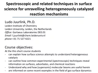
Spectroscopic and related techniques in surface science for unravelling heterogeneously catalyzed reaction mechanisms • Ludo Juurlink, Ph.D. • Leiden Institute of Chemistry • Leiden University, Leiden, the Netherlands • Office:Gorlaeus Laboratories DE0.01 • Email: l.juurlink@chem.leidenuniv.nl • phone +31 71 527 4221 • Course objectives: • At the this short course students • can explain how surface science attempts to understand heterogeneous catalysis • can outline how common experimental (spectroscopic) techniques reveal information on surfaces, adsorbates, and chemical reactions • Understand why and how (supersonic) molecular beams are useful herein • are informed on some recent examples in the field of gas-surface dynamics
Electron spectroscopy • Probing of electronic structure of the surface through analysis of energy of secondary electrons emitted from sample. • Auger electron spectroscopy (AES) • Determines: surface composition • Uses: irradiation via electrons • Photoelectron spectroscopy (XPS, UPS) • Determines: surface composition and electronic structure • Uses: irradiation via photons
Elastic peak electrons photons ions secondary electrons 0 Ep N(E) Spectrum of secondary electrons N(E) Ep Ep = 5-3000 eV with a narrow Ep
electrons photons ions secondary electrons N(E) Spectrum of secondary electrons Ep Ep = 5-3000 eV with a narrow Ep
Electron energy analyzers • 4-grid LEED optics: retarding field analyzer • electrons with E0<eV0 are repelled • applying a sine wave to the potential ramp with LIA detection enhances sensitivity
Electron energy analyzers • Cylindrical mirror analyzer (CMA): deflection analyzer • electrons in narrow energy window are detected • applying a sine wave to the potential ramp with LIA detection enhances sensitivity
Electron energy analyzers • Concentric hemispherical analyzer (CHA): deflection analyzer • electrons in narrow energy window are detected
Lise Meitner 1878-1968 Pierre Auger 1899-1993 Auger electron spectroscopy Uses an electron beam to create an initial state of a hole in a (surface) atom. Through the Auger process, a second hole is created and a second electron emitted with a specific Ekin. Surface composition can be determined as every atom has unique atomic energy levels.
Auger electron spectroscopy Ni(cyl) O C S Dirty Cu sample
Auger electron spectroscopy • Quantitative analyses are possible for > 0.01 monolayer of adsorbateor alloy • A standard is required (e.g. an adsorbate yielding a known maximum coverage)
Lise Meitner 1878-1968 Pierre Auger 1899-1993 Auger electron spectroscopy Uses an electron beam to create an initial state of a hole in a (surface) atom. Through the Auger process, a second hole is created and a second electron emitted with a specific Ekin.
Photoelectron spectroscopy • Based on photoelectric effect: electron with binding energy Eiabsorbs photon with energy ħω, and leaves solid with kinetic energy: Ekin=ħω - Ei – φ • where φ = Evacuum-EFermiis the work function of the material. • Conditions to detect escaping electron: • ħω > Ei + φ • Electron velocity is directed towards outer surface • Electron does not loose energy due to collisions with other electrons on its way to the surface
X-ray source or UV lamp Laboratory photoelectron spectroscopy Ekin=ħω - Ei – φ • X-ray photoelectron spectroscopy (XPS) • Mg K1,2ħω =1253.6 eV (9.891 Å) • Al K1,2ħω =1486.6 eV (8.341 Å) • Ultraviolet photoelectron spectroscopy (UPS) • discharge lamp He: ħω = 21.22 eV (58.43 nm) • 40.81 eV (30.38 nm) Alternative: synchrotron facility
X-ray photoelectron spectroscopy X-ray photoelectron spectroscopy Ei=ħω - Ekin- φ
X-ray photoelectron spectroscopy X-ray photoelectron spectroscopy: Core level shifts Due to different environment of surface versus bulk atoms
X-ray photoelectron spectroscopy X-ray photoelectron spectroscopy: Core level shifts Chemical changes lead to large shifts. C2H4/Co(0001) C C2H2 C2H4 Courtesy of: C.J. Weststrate
Ultraviolet photoelectron spectroscopy • Ultraviolet photon spectroscopy (UPS): low photon energies (< 50 eV), so only valence levels become excited • Angle-integrated UPS • Angle-resolved UPS (ARUPS)
Vibrational spectroscopies (High Resolution) Electron Energy Loss Spectroscopy (HREELS) and Reflection Absorption Infrared Spectroscopy (RAIRS)
Electron energy loss spectroscopy • Study of inelastically scattered electrons, which have lost well-defined energies during interaction with surface • Different scattering processes: • Core level excitation (CLEELS): 100 – 104eV • Excitation of plasmons and electronic interband transitions (EELS): 1 – 100 eV • Excitation of vibrations of surface atoms and adsorbates (HREELS): 10-3 – 1 eV • Scattering mechanisms • Dipole scattering – long range • Very sharp features near specular angle • Impact scattering – short range • Wide angle scattering • Negative ion resonances • Strong dependence on impact energy e- Ekin,in Ekin,out
Electron energy loss spectroscopy • HREELS: • Identification of adsorbed species • Identification of adsorption sites • Identification of spatial orientation of adsorbed molecule
Electron energy loss spectroscopy • HREELS: • Identification of adsorbed species • Identification of adsorption sites • Identification of spatial orientation of adsorbed molecule
Reflection Absorption InfraRed Spectroscopy (RAIRS) • Highest sensitivity for observing an absorption feature when • p-polarized light • grazing incidence • molecule with transition dipole arranged along surface normal • molecule with large transition moment Selection rule:
Reflection Absorption InfraRed Spectroscopy (RAIRS) Chen et al., Faraday Discuss. 157, 285 (2012)
Reflection Absorption InfraRed Spectroscopy (RAIRS) 50 ML Amorphous Solid Water Crystalline ice 1 ML ASW on H/Pt(533) free OH group
Temperature Programmed Desorption (TPD) or Thermal Desorption Spectroscopy (TDS) or Temperature Programmed Reaction Spectroscopy (TPRS)
physisorption dissociative chemisorption Potential energy diagram O2/M(hkl) molecular chemisorption • The depth of the invidual well depends on • material • surface structure • adsorption site • molecular orientation Dissociation energy in the gas phase O2 (g) 2 O(g) V(r) O2, chem The crossings of the adsorption curves determine whether a process is activated or not. O2, phys 0 O2 (g) adiabatic behavior Oads + Oads r
Potential energy surface O2/Pt(111) from: Groß, Eichler, Hafner, Mehl, and Papaconstantopoulos, Surf. Sci. 539, L542 (2003)
Rate of adsorption: Rate of desorption: Desorption Desorption is the reverse process of adsorption • Molecular or atomic desorption • Recombinative desorption (opposite of dissociative adsorption) V(r) A2 (g) 0 A2, phys A2,chem occupied fraction of the surface r 2 Achem
Rate of adsorption: Rate of desorption: Desorption Desorption is the reverse process of adsorption • Molecular or atomic desorption • Recombinative desorption (opposite of dissociative adsorption) V(r) A2 (g) 0 A2, phys A2,chem r 2 Achem
Langmuir isotherm Langmuir adsorption model • Only one type of adsorption site • All these sites are equivalent • Only one adsorbate per site/no interactions At equilibrium rates add up to zero. non-dissociative dissociative
Temperature Programmed Desorption The experiment • cool sample under UHV conditions • expose it to the relevant gas • heat the sample while monitoring desorption • Quadrupole Mass Spectrometer • track Aads spectroscopically • … Polanyi-Wigner equation and Note:
Example H2O interaction with Pt[n(111)x(100)]
Temperature Programmed Desorption H2O/Pt(111) From: Hay et al., Surf. Sci. 505, 171 (2002)
Temperature Programmed Desorption H2O/D/Pt(S) H2O/Pt(S) Pt(111) Pt(533) Pt(755) Pt(977) From: Den Dunnen et al., PCCP 17, 8530 (2015)
Temperature Programmed Desorption D2/Cu(211) vs D2/Cu(111) From: Kao, Kleyn and Juurlink, in preparation
Temperature Programmed Desorption D2/Cu(211) vs D2/Cu(111) From: Kao, Kleyn and Juurlink, in preparation
Temperature Programmed Desorption D2/Cu(211) vs D2/Cu(111) From: Kao, Kleyn and Juurlink, in preparation