Download
1 / 89

E N D
Shulman and Rothman PNAS, 1998 In this period of intense research in the neurosciences, nothing is more promising than functional magnetic resonance imaging (fMRI) and positron emission tomography (PET) methods, which localize brain activities. These functional imaging methodologies map neurophysiological responses to cognitive, emotional, or sensory stimulations. The rapid experimental progress made by using these methods has encouraged widespread optimism about our ability to understand the activities of the mind on a biological basis. However, the relationship between the signal and neurobiological processes related to function is poorly understood, because the functional imaging signal is not a direct measure of neuronal processes related to information transfer, such as action potentials and neurotransmitter release. Rather, the intensity of the imaging signal is related to neurophysiological parameters of energy consumption and blood flow. To relate the imaging signal to specific neuronal processes, two relationships must be established… The first relationship is between the intensity of the imaging signal and the rate of neurophysiological energy processes, such as the cerebral metabolic rates of glucose (CMRglc) and of oxygen (CMRO2). The second and previously unavailable relationship is between the neurophysiological processes and the activity of neuronal processes. It is necessary to understand these relationships to directly relate functional imaging studies to neurobiological research that seeks the relationship between the regional activity of specific neuronal processes and mental processes.
Shulman and Rothman PNAS, 1998 Psychology Image Signal Mental Neuroenergetics Neuronal CMRglc CMRO2 CBF Neuroscience
Hemoglobin Molecule 280 million Hb molecules per red blood cell
Different magnetic properties of hemoglobin and deoxyhemoglobin L. Pauling and C. Coryell The Magnetic Properties and Structure of Hemoglobin, Oxyhemoglobin and Carbonmonoxy hemoglobin, PNAS, vol. 22, pp. 210-216, 1936.
Blood Oxygenation Level Dependent Imaging Baseline Task from Mosley & Glover, 1995
Virchow-Robin Space Large Vessel Contributions to BOLD Contrast
Intravascular Perivascular Extravascular
3 z = 1.64 Large Small Isotropic Diffusion Weighted Spiral Imaging at 4T Courtesy of Dr. Allen Song, Duke University
a b 9 sec 9 sec
BOLD activation (b factor = 0) Diffusion-weighted (b factor = 54) Diffusion-weighted (b factor = 108) ADC masked by BOLD activation Subject 41057, Slice 12, 4.0 Tesla
BOLD activation (b factor = 0) Diffusion-weighted (b factor = 54) Diffusion-weighted (b factor = 108) ADC masked by BOLD activation Subject 41037, Slice 183, 4.0 Tesla
BOLD activation (b factor = 0) Diffusion-weighted (b factor = 54) Diffusion-weighted (b factor = 108) ADC masked by BOLD activation Subject 41037, Slice 177, 4.0 Tesla
BOLD activation (b factor = 0) ADC masked by BOLD activation Subject 41037, Slice 177, 4.0 Tesla
Phosphorescence Decay Time (Oxyphor R2 oxygen tension-sensitive phosphorescent probe) Vanzetta and Grinvald, Science, 286: 1555-1558, 1999
Phosphorescence Decay Time (Oxyphor R2 oxygen tension-sensitive phosphorescent probe) Vanzetta and Grinvald, Science, 286: 1555-1558, 1999
Vanzetta and Grinvald, Science, 286: 1555-1558, 1999 deoxy Hb Oxy Hb
Berwick et al, JCBFM, 2002 Optical imaging of rat barrel cortex Hb02= oxyhemoglobin, Hbr = deoxyhemoglobin, Hbt = total blood flow
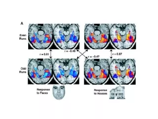
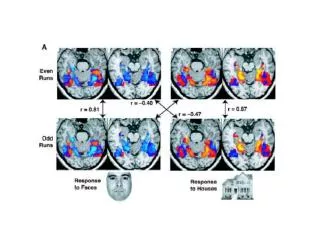
Functional Imaging of the Monkey Brain N. Logothetis, Nature Neuroscience, 1999
Early Response in fMRI Hu, Le, Ugurbil MRM, 1997
Early Response in fMRI Hu, Le, Ugurbil MRM, 1997
Arterioles (10 - 300 microns)precapillary sphinctersCapillaries (5-10 microns)Venules (8-50 microns)
Tissue factors • K+ • H+ • Adenosine • Nitric oxide
Neuronal Control of the Microcirculation C. Iadecola, Nature Neuroscience, 1998 Commentary upon Krimer, Muly, Williams and Goldman-Rakic, Nature Neuroscience, 1998
Pial Arteries Noradrenergic Dopamine 10 m Krimer, Muly, Williams, Goldman-Rakic, Nature Neuroscience, 1998
Dopamanergic terminals associated with small cortical blood vessels 10 m Krimer, Muly, Williams, Goldman-Rakic, Nature Neuroscience, 1998
Dopamanergic terminals associated with small cortical blood vessels 2 m 400 nm 2 m 400 nm Krimer, Muly, Williams, Goldman-Rakic, Nature Neuroscience, 1998
Perivascular iontophoretic application of dopamine 18-40 s 40-60 s Krimer, Muly, Williams, Goldman-Rakic, Nature Neuroscience, 1998
glucose glucose Glucose 6 phosphate Net +2 ATP Fructose – 1,6-phosphate pyruvate lactate TCA cycle O2 Net +36 ATP CO2 + H20
Shulman and Rothman PNAS, 1998 Proposed pathway of glutamate / glutamine neurotransmitter cycling between neurons and glia, whose flux has been quantitated recently by 13C MRS experiments. Action potentials reaching the presynaptic neuron cause release of vesicular glutamate into the synaptic cleft, where it is recognized by glutamate receptors post-synaptically and is cleared by Na+ -coupled transport into glia. There it is converted enzymatically to glutamine, which passively diffuses back to the neuron and, after reconversion to glutamate, is repackaged into vesicles. The rate of the glutamate-to-glutamine step in this cycle (Vcycle), has been derived from recent 13C experiments.
Attwell and Laughlin, JCBFM, 2001 Brain Energetics
Attwell and Laughlin, JCBFM, 2001 Brain Energetics
Lauritzen, JCBFM, 2001 Climbing Fiber Stimulation