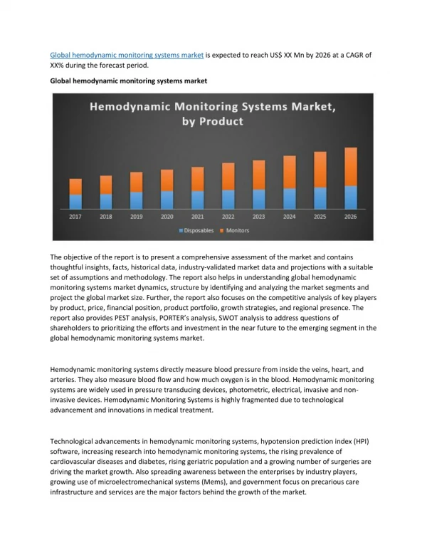
Download
1 / 21
230 likes | 1.17k Vues
Hemodynamic Monitoring and Transthoracic Lines. Deb Updegraff RN, CNS Lucille Packard Children’s Hospital Pat Hock RN, Nurse Educator. Winnie Yung , CNS. Infants and children undergoing open heart surgery may require intracardiac monitoring.

E N D
Hemodynamic Monitoring and Transthoracic Lines Deb Updegraff RN, CNS Lucille Packard Children’s Hospital Pat Hock RN, Nurse Educator Winnie Yung , CNS
Infants and children undergoing open heart surgery may require intracardiac monitoring. The hemodynamic data can assist in the assessment of contractility, preload and afterload. As the patient stabilizes post cardiac by-pass, intracardiac catheters (RA) may be left in place for vascular access reasons.
What’s the difference ?? “Percutaneous” vs “Transthoracic” Percutaneous – Insertion site is through the skin. Transthoracic- Insertion is done while the chest is open and directly through the myocardium.
Examples of Percutaneous lines: • PICCs • Tunneled lines • Non-tunneled lines • Swan-Ganz thermodilutional catheters • Dialysis/CRRT catheters
Percutaneous Central Venous Catheter PA Pulmonary Artery Catheter LA Left Atrial Transthoracic Catheter RA Right Atrial Transthoracic Catheter Roth, S. 1998
Right Atrial Pressure MonitoringIndications • Measure right atrial pressure (RAP) • Same as Central Venous Pressure (CVP) • Assess blood volume; reflects preload to the right side of the heart • Assess right ventricular function • Infusion site for large fluid volume • Infusion site for hypertonic solutions
Reasons for elevated RA pressure: • decreased right (or single) ventricle compliance • tricuspid valve disease • Intravascular volume overload • cardiac tamponade • tachyarrhythmia • Right Atrial Pressure • Mean: 1 to 7 mm Hg
Reasons for reduced RA pressure: • low intravascular volume status • inadequate preload • Right Atrial Pressure • Mean: 1 to 7 mm Hg
Pneumothorax Hemothorax Hemorrhage Cardiac tamponade Vessel, RA, or RV perforation Arrhythmias Air embolism Pulmonary embolism Thromboembolism Infection Right Atrial Pressure MonitoringComplications
Right Atrial Pressure MonitoringWaveform Analysis • a wave: rise in pressure due to atrial contraction • x decent: fall in pressure due to atrial relaxation • c wave: rise in pressure due to ventricular contraction and closure of the tricuspid valve • v wave: rise in pressure during atrial filling • y decent: fall in pressure due to opening of the tricuspid valve and onset of ventricular filling
Elevated RAP RV failure Tricuspid regurgitation Tricuspid stenosis Pulmonary hypertension Hypervolemia Cardiac tamponade Chronic LV failure Ventricular Septal Defect Constrictive pericarditis Decreased RAP Hypovolemia Increased contractility Right Atrial Pressure MonitoringWaveform Analysis
Reasons for elevated LA pressure: • Elevated systemic ventricular end diastolic pressure • mitral valve disease • Large left-to-right shunt • intravascular volume overload • cardiac tamponade • tachyarrhythmia • Artifactual
Reasons for reduced LA pressure: • low intravascular fluid status • Inadequate preload • Artifactual
Reasons for elevated PA pressure: • mechanical obstruction of pul. circulation • pul. arteriolar smooth muscle hypertrophy • inflammatory response to CPB • mechanical obstruction of the airways (for examples…) • acidosis and hypoxia • elevated LA pressure • unrestrictive VSD or large PDA • pul. hypertension
Nursing HOURLY assessment: • Air in line or stopcocks • Precipitates • Leaking at site • Increasing resistance • Condition of entrance sites
BEFORE REMOVAL Transthoracic Line • Check coagulation labs (pt, ptt, INR, platelets) • Transfuse if Platelets < than 70 and INR > 1.5 • Ensure Packed Red Blood Cells in cooler at bedside • (Remember two RN check for PRBCs. Instructions • for blood in cooler, taped to cooler) • Ensure good vascular access • Ensure chest tube patency • Evaluate need for sedation. (if too active ↑ BP may → bleeding)
After Removal of Transthoracic Line • Keep PRBCs for a minimum of 1 hour • Continuous hemodynamic monitoring for a minimum of 1 hour • (assess for signs of tamponade-dampening arterial wave form • narrowing pulse pressure and bleeding- blood in chest tubes, • decrease blood pressure, pallor • altered LOC) • Document vitial signs every 15 minutes • Check HCT if bleeding suspected • Ensure patency of chest tubes • Do not transfer patient for at least 2 hours
Pressure Line Safety • What is air vigilance and why is it so important? • Why is it unsafe to draw back or flush fluid into a line infusing vasoactive medications? • What precautions should be taken when discontinuing any pressure line? • Is it safe to get a patient out of bed to be held or to sit in a chair if they have a transthoracic pressure line? • What additional safety measures should be followed for transthoracic pressure lines?
References • Alspach. AACN’s Core Curriculum for Critical Care Nursing. Saunders. • Berne and Levy. Physiology. Mosby. • Hazinski. Manual of Pediatric Critical Care. Mosby. • Kinney, Packa, and Dunbar. AACN’s Clinical Reference for Critical Care Nursing. Saunders. • Kumm. Hemodynamic Monitoring. University of Kansas School of Nursing. • Kumm. Intra-arterial Pressure Monitoring. University of Kansas School of Nursing. • Slota. AACN’s Core Curriculum for Pediatric Critical Care Nursing. Saunders. • Taleghani, Fred. Invasive lines, hemodynamic monitoring, and waveforms. LPCH, PICU.