Download
1 / 6
70 likes | 247 Vues
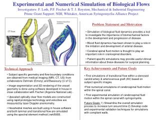
Experimental and Numerical Simulation of Biological Flows Investigators: F. Loth, P.F. Fischer & T. J. Royston, Mechanical & Industrial Engineering Prime Grant Support: NIH, Whitaker, American Syringomyelia Alliance Project. Problem Statement and Motivation.

E N D
Experimental and Numerical Simulation of Biological Flows Investigators: F. Loth, P.F. Fischer & T. J. Royston, Mechanical & Industrial Engineering Prime Grant Support: NIH, Whitaker, American Syringomyelia Alliance Project Problem Statement and Motivation • Simulation of biological fluid dynamics provides a tool to investigate the importance of biomechanical factors in the development and progression of disease. • Blood fluid dynamics has been shown to play a role in the initiation and development of arterial disease. • Cerebral spinal fluid motion is thought to play an important role in craniospinal disorders. • Patient specific simulations may provide useful clinical information about these diseases for surgical planning. Key Achievements and Future Goals Technical Approach • Subject specific geometry and flow boundary conditions are obtained from medical imaging (MRI, CT, US) from collaborators Oshinski (Emory) and Bassiouny (U of C). • Image segmentation and 3D rendering of the vessel geometry is done using software developed in house in close collaboration with Fischer (Argonne National Lab) . • Upscaled optically clear flow models are constructed using rapid prototype technology and velocities are measured by laser Doppler anemometry. • Hexahedral meshes are built using in house software and both laminar and transitional flow are simulated using the spectral element method (nek5000). • First simulations of transitional flow within a stenosed carotid artery & arteriovenous graft (AV) based on subject specific images. • First numerical simulations of cerebrospinal fluid motion within the spinal canal. • First experimental simulation of cerebrospinal fluid motion within the spinal canal with syringomyelia • Future Goals: 1) Streamline the overall simulation process to increase turn around time 2) Develop code and experimental validation techniques for simulations with compliant walls.
Multimode Sonic & Ultrasonic Diagnostic Imaging Investigators: Thomas J. Royston & Francis Loth, Mechanical & Industrial Engineering Prime Grant Support: NIH Artery Fluid flow Noise generation Approximate location of constriction Problem Statement and Motivation Bimodal image. • Ultrasonic (US) imaging provides detailed geometry • Geometric changes may indicate disease or injury • Sonic imaging provides unique functional information • Sounds associated with disease are sonic, not US • Merge US and Sonics to harness strengths of each • Initial application: peripheral vascular pathologies – vessel constrictions (plaque and intimal hyperplasia) Blood vessel with constriction in soft tissue phantom Grayscale of geometry from US imaging Color overlay of acoustic field generated by turbulence downstream of the constriction Key Achievements and Future Goals Technical Approach • Sonic wave propagation in biological tissue is more complex than US. • Requires new acoustic modeling developments • Inverse modeling to extract acoustic image from array • Novel acoustic sensor development • Prototype US/Sonic system has been developed • - conventional US system retrofitted with • - electromagnetic position device for true 3D imaging • - acoustic sensor array pad that is transparent to US so US imaging can be conducted with the pad in place • Calibration of system on phantom models in progress • Turbulence imaged downstream of vessel constriction • Future plans: Human subject studies, improved prototype, better sensor array, improved imaging software Prototype 15 sensor sonic array pad on arm • Merging multiple imaging modalities on same platform Biomedical & Biotechnology
Biomimetic MEMS Technology for a Novel Retinal Prosthesis PI: Laxman Saggere, Mechanical and Industrial EngineeringCollaborator: David Schneeweis, BioEngineering Prime Grant Support: National Science Foundation Problem Statement and Motivation • Motivation: Photoreceptor degeneration in diseases such as ARMD and RP is the leading cause of blindness in the world. No cures or therapies are available for these diseases, but a retinal-based prosthesis offers a promising treatment option. Most current retinal prostheses rely on the concept of electrical stimulation of neurons, which is conceptually simple, but faced with many challenges • Objective: To develop a biomimetic technology enabling a fundamentally different and technically superior approach to a retinal prosthesis. This approach, in principle, mimics a natural photoreceptor’s function of transducing visual stimuli into chemical signals that stimulate the surviving retinal neurons. Array of light-powered microactutor prototypes Technical Approach Key Achievements and Future Goals • Challenges: i)Low intensity light at the retina; ii) Integration of array components and microfluidics; iii) Chemical dispensing rate, mechanism, long-term operation; iv) Biocompatible packaging. • Key Achievements: i) Completed preliminary system design and established the concept feasibility; ii) Established a technique to chemically stimulate neuronal cells and record the cellular response; iii) Fabricated and characterized the light powered actuator; iv) Established techniques to quantify nanoliter flow • Future Goals: i) To fabricate and test an in-vitro proof of the concept device; ii) To lead the technology developed towards clinical relevancy through interdisciplinary collaborations with neuroscientists and retina specialists. • Approach: A microdispenser unit integrated with a miniaturized solar cell and a thin-film piezo actuator on one side and several micron-scale ports on the other side contains liquid chemical (neurotransmitter). An array of such microdispenser units constitutes the core of a prosthesis. • Principle of Operation: Light falling on the retina irradiates the solar cell, which generates voltage across the piezo actuator. The actuator pressurizes the liquid and dispenses it through the micro ports. The liquid diffuses through micro-capillaries in a soft encapsulation and stimulates retinal cells. • Technologies: MEMS, microfluidics, thin-film piezoelectric actuators, solid-sate solar cells, chemical cellular signaling. MIE – Biotechnology and Micro/Nano Emphasis Areas
Effects Of Bone Mineral Density And Surgical Technique On Stability Of Acetabular Cup After Total Hip Replacement Investigators: Ivan Zivkovic1; Farid Amirouche1; Mark Gonzalez2 1Department of Mechanical Engineering and 2Department of Orthopedic Surgery Prime Grant Support: Zimmer Orthopedic Problem Statement and Motivation • Total hip replacement surgery has become a common procedure to alleviate pain caused by osteoarthritis, rheumatoid arthritis, fractures, and other hip related problems for patients over 55 years of age. • With the aging of the global population, the demand for hip replacements is increasing, along with the required clinical lifetime. • The goal of this research is to study the effect of aging and surgical technique on stability of a hip prosthesis and ultimately to improve durability of hip joint prosthesis. Technical Approach • Experimental cadaveric study was conducted to measure initial relative micromotion at the prosthesis/bone interface and to investigate the effect of bone density and surgical technique on the early micromotion at the interface that may predispose to a prosthesis loosening. • Sensor technology was used to capture the micromotion of acetabular prosthesis • Image-processing package (SeScan 3.0) was designed to generate a 3-D bone geometry and material distribution from ST scan and MRI data. • Parametric patient based finite element model, validated with experimental results, was developed to further analyze the conditions affecting the initial stability and loosening of the interface for different loading conditions. Key Achievements and Future Goals • Patient specific computer system is developed which couples clinical imaging with finite element method • This increased interpretive power has the potential to streamline biomedical diagnosis, analysis, non-invasive surgical planning and most importantly computer-assisted surgery • At the initial clinical consultation proposed system would warn orthopedic surgeon of any anatomical abnormalities that could jeopardize the implant fixation, helps in determining optimal positioning of the prosthesis, insertion method, etc. which leads to reduction of operating time and to enchased patient care.
Orienting Human Stem Cells (hMSCs) by Means of Electrospun Polymer Nanofibers Investigators: M. Cho, Bioengineering; A. Yarin, C. M. Megaridis, Mechanical and Industrial Engineering; E. Zussman, Technion-Israel Oriented Random Problem Statement and Motivation • Cell orientation and adhesion control the functionality of natural and engineered tissues • Electrospinning is a low-cost technique which can produce polymer nanofibers aligned along a specific direction • Polymer nanofibers can be used to mimic the native extracellular matrix (ECM) features • Electrospun polymer nanofiber scaffolds are used to manipulate cell orientation and adhesion Cells: Green, Nanofibers: Red Key Achievements and Future Goals Technical Approach • Random and oriented polycaprolactone (PCL) nanofibrous scaffolds produced using electrospinning • hMSCs were cultured and seeded on two scaffold types (random, oriented) • Orientations of hMSCs and nanofibers on random and oriented nanofibrous scaffold samples were measured via laser scanning confocal microscopy at different time points during an 18-day culture period • hMSC viability tests were performed to verify compatibility of the cells with the PCL • hMSCs adhered and oriented along PCL nanofibers • During long-term culture, hMSCs demonstrated no preferred orientation on random nanofibrous scaffolds; cells consistently aligned on oriented scaffolds • Oriented PCL nanofibrous scaffolds could be used to mimic the cell and ECM organization in the native tissue, such as muscle, tendon, and the superficial zone of articular cartilage • The fiber scaffold/hSMC approach holds promise for a variety of tissue engineering applications
Multi-scale Modeling of Failure in Cortical Bone Investigator: Elisa Budyn, Mechanical Engineering Grant Support: UIC; Collaboration: Ecole Centrale Paris (Thierry Hoc, Material Science) Problem Statement and Motivation • Determination of the effects of the local geometrical and material heterogeneities in sane and pathological cortical bone at the micro and nano scales over the local strain and stress fields and global response of the unit cells. • A better understanding of the effect of pathologies over cortical bone quality Key Achievements and Future Goals Technical Approach • Multi-scale numerical models to characterize the mechanics of materials and biomaterials with multi-phase complex microstructures. • Failure mechanics of these microstructures though damage and fracture processes studied over the micro and nano scales, modeled through FEM and X-FEM approaches. • Concomitant experiments over the multiple scales. • Determination of the RVE • Determination of the Macroscopic Moduli • Effect of the cement lines over the local strain field and the work of separation due to crack propagation • Determination of localization patterns • Crack initiation and crack propagation in cortical bone