Download

1 / 9
0 likes | 1 Vues
Liver ischemia reperfusion is induced during sur-gical procedures like liver transplantation and re-section. Multiple mechanisms have been postulat-ed to liver damage following liver ischemia reperfu-sion injury, such as oxidative stress and inflamma-tory reactions. The present study declares the pos-sible mechanism of tadalafil, toward modulating the inflammatory response. Forty-eight rats were divided into 4 groups as follows; Sham group sub-jected to midline laparotomy only. Tadalafil group administered Tadalafil 10 mg/kg intraperitoneal 45 min before sham operation. I/R (Ischemia-reperfusi

E N D
Eur. J. Anat. 23 (5): 325-332(2019) ORIGINAL ARTICLE Liver ischemia/reperfusion injury, a setting in which the functional mass is reduced and the role of PDE5 inhibitor Hesham N. Mustafa1, Gehan A. Hegazy2,3, Sally A. El Awdan4, Aliaa Amr Alamoudi2 1Anatomy Department, Faculty of Medicine, King Abdulaziz University, Jeddah, KSA, 2Clinical Biochemistry Depart- ment, Faculty of Medicine, King Abdulaziz University, Jeddah, KSA, 3Medical Biochemistry Department, National Re- search Centre, Cairo, Egypt, 4Pharmacology Department, National Research Centre, Cairo, Egypt SUMARY Liver ischemia reperfusion is induced during sur- gical procedures like liver transplantation and re- section. Multiple mechanisms have been postulat- ed to liver damage following liver ischemia reperfu- sion injury, such as oxidative stress and inflamma- tory reactions. The present study declares the pos- sible mechanism of tadalafil, toward modulating the inflammatory response. Forty-eight rats were divided into 4 groups as follows; Sham group sub- jected to midline laparotomy only. Tadalafil group administered Tadalafil 10 mg/kg intraperitoneal 45 min before sham operation. I/R (Ischemia- reperfusion) group, rats undergo 60 min of hepatic ischemia followed by 60 min of reperfusion. Tada- lafil + I/R group rats undergo a similar pattern of I/ R after the treatment with Tadalafil 10 mg/kg, 45 min before ischemia. At the end of the reperfusion, the blood samples were collected for estimation of biochemical markers including liver enzymes using colorimetric assay method and serum: TNF-α (tumor necrosis factor-α), IL-6 (interleukin 6) le- vels, ICAM- 1 (Intercellular Adhesion Molecule-1) were measured. Tissues were evaluated by semi- quantitative and morphometrical approaches. Ta- dalafil succeeded in restoring normal levels of liver enzymes and ameliorating the oxidative stress as evidenced by decreasing MDA and restoring redu- ced glutathione levels in liver tissue homogenate. Also, Tadalafil exhibits anti-inflammatory effects, as it significantly decreased the levels of TNF-α, IL6 and ICAM-1. The findings are supported by BCL-2, TNF-α immunomarkers. It is concluded that modulation of the inflammatory response might be one of the mechanisms of Tadalafil-mediated he- patoprotection, so it is recommended as an adju- vant therapy in liver surgery. Key words: Ischemia/reperfusion injury – Oxidati- ve stress – Apoptosis – TNF-α –BCL-2 INTRODUCTION Hepatic I/R (ischemia/reperfusion) injury is a common complication following surgical procedu- res such as liver resection and transplantation that involves a variable period of ischemia, and may result in complicated medical conditions such as shock, trauma, or low cardiac output (Ye et al., 2015). In addition, it may occur during liver surgery when Pringle’s maneuver (ligation of the hepato- duodenal ligament) to reduce blood loss is done Authors’ contributions: All authors participated in the design of this work and made equal contributions. All authors read and Corresponding author: Dr. Hesham N. Mustafa, M.D. Depart- approved the final manuscript ment of Anatomy, Faculty of Medicine, King Abdulaziz Universi- ty, P.O. Box 80205, Jeddah 21589, Saudi Arabia. E-mail: hesham977@hotmail.com Submitted: 25 March, 2019. Accepted: 3 June, 2019. 325
Liver ischemia/reperfusion injury and PDE5 inhibitor role (Freitas et al., 2017). Ischemia initiates a series of events that cause necrosis and cellular dysfunction; the reperfusion of blood flow can paradoxically generate more tis- sue injury. Excessive inflammatory reaction is con- sidered as a key mechanism. A complex cascade of inflammatory mediators is activated by liver ischemia and reperfusion (Zhai et al., 2011). I/R injury is mediated by the activation of proinflamma- tory cytokines such as TNF-α (tumor necrosis fac- tor alpha), release of free radicals and the accu- mulation of inflammatory cells, accumulation of intracellular sodium and calcium, and induction of hepatocyte apoptosis (Rao et al., 2013). During hepatic I/R injury, many microvascular and endothelial alternations occur as initiation of coagulation cascade, variations in the molecular vasoactive products such as endothelin (ET) and NO (nitric oxide), and upregulation of endothelial adhesion molecules such as ICAM-1 (intracellular adhesion molecule-1). These alternations are sug- gested targets for therapeutic strategies (Camara- Lemarroy et al., 2014). Safe clearance of damaged cells is the apoptosis (programmed cell death). BCL-2 family are proteins that are considered as a non-death signal (antiapoptosis) (Hegazy et al., 2018). Phosphodiesterase (PDE) inhibitors are the com- pounds that inhibit the biosynthesis or actions of PDEs. PDE inhibitors are currently widely used in treatment of erectile dysfunction, as well as in pul- monary arterial hypertension (Sawamura et al., 2009). PDE inhibitors have protective effects on myocardial muscles and vascular structures (Korkmaz-Icoz et al., 2018). The effects of PDE inhibitors have been widely studied in I/R in diffe- rent organs. Moreover, PDE is the family of enzy- mes that adjust the cellular levels of second mes- sengers, cyclic adenosine monophosphate (cAMP) and cyclic guanosine monophosphate (Reffelmann and Kloner, 2009). Tadalafil (TDF) is a selective and effective inhibi- tor of PDE5 (phosphodiesterase type-5) that has been broadly used in the treatment of erectile dys- function due to its capability to prevent the break of cGMP, which is the second messenger of NO (nitric oxide) (Kucuk et al., 2012). The purpose of this study was to declare the beneficial effect of tadalafil on hepatic I/R-injury. MATERIALS AND METHODS Drugs Tadalafil was purchased from Lilly Co. and given via intraperitoneal injection with dose of 10 mg/kg (Bektas et al., 2016). Animals Mature male Wistar albino rats, their weighs ran- ging from 200 to 250g. They were obtained from the Animal House. They were preserved in cages with optimum temperature, 60% humidity under 12h dark and light cycles. Rats were provided standard pellet diet and water for one week before the experiment for adaptation. Ethics Statement This study was approved by the biomedical Ethics Research Committee [Reference No 229- 19]. Experimental Design Forty-eight rats were separated into four groups (12 each) in this way; Sham group underwent midline laparotomy only. Tadalafil group administe- red Tadalafil 10 mg/kg intraperitoneal 45 min befo- re sham operation (Bektas et al., 2016). Ischemia- reperfusion (I/R) group, rats exposed to 60 min of hepatic ischemia, then they were exposed to 60 min of reperfusion (Liu et al., 2016). Tadalafil+ I/R group rats were subjected to a similar pattern of I/ R after the administration of Tadalafil 10 mg/kg, 45 minutes before ischemia (Bektas et al., 2016). Surgical Procedure All surgical procedures were performed under complete aseptic conditions, and the anesthesia, with combination of ketamine 100 mg/kg and xyla- zine 10 mg/kg, administered through intraperito- neal route (Savvanis et al., 2014). Rats were expo- sed to midline abdominal incision, liver lobes and the portal triad identified, and rats subjected to 60 minutes of hepatic ischemia by clamping the portal triad with a micro-vascular clamp after the bifurca- te of the right lobe, interrupting the portal triad flow to the left and median lobes, inducing ischemia for 60 min, and after that the clamp removed to allow 60 min of reperfusion (Hueper et al., 2018). At the end of the reperfusion period, the blood samples were collected from the abdominal aorta and biop- sies were taken from the ischemic hepatic lobes. Collection for blood samples Blood was centrifuged (700×g, 4°C, 20 min) for assessment of liver enzymes. Liver tissue Extracts Liver was homogenized in phosphate buffer sali- ne (PBS) [10%]. The first part was centrifuged at 15000 g for 10 min, and the supernatant was gat- hered and kept at -80C° to measure oxidative stress markers and inflammatory cytokines in liver tissue homogenates. The second part was subjec- ted to repeated freeze-thaw cycle twice in order to break the cell membranes, then centrifuged at 5000×g for 5 min and kept at -80C° for the measu- rement of the other parameters. Hepatic biochemical parameters in serum Serum ALT and AST were determined by using colorimetric assay kits provided by Elabscience, Houston, Texas, USA, (Catalogues E-BC-K235, E- 326
H. N. Mustafa et al. Table 1: Effect of Tadalafil on serum liver functions. Sham N=12 Tadalafil N=12 304.33±57.90 1P= 0.686 2P= 0.000 268.83±50.59 1P= 0.863 2P= 0.000 I/R N=12 Tadalafil +I/R N=12 299.75±.53.26 1P= 0.809 2P= 0.000 378.25±181.79 1P= 0.018 2P= 0.000 Groups 491.75±85.35 1P= 0.000 ALT (U/L) 293.00±71.39 791.25±126.37 1P= 0.000 AST (U/L) 260.50±58.77 Values are Mean± SD: standard deviation. Comparison between groups done by ANOVA followed by LSD post hoc test. 1P: compared to Sham group. 2P: compared to I/R (group subjected to ischemia reperfusion injury). ALT (Alanine transaminase), AST (Aspartate transaminase). U/L unit/ liter. BC-K236 respectively) in accordance with the ma- nufacturer’s instructions. Oxidative stress markers activities in liver tis- sue homogenates Estimation of MDA (malondialdehyde) and GSH (reduced glutathione) levels utilizing colorimetric assay kits (Catalogues No.MD 25 29, GR 25 11 respectively) according to the manufacturer’s ins- tructions (Bio Diagnostic, Cairo, Egypt). Assessment of hepatic cytokines in liver tissue homogenates TNF-α (Tumor necrosis factor-α) and IL-6 (interleukin 6) levels were measured using ELISA (enzyme-linked immunosorbent assay) kit; (TNF-α: catalogue number RAB0479 Sigma), RayBio Rat IL-6 ELISA Kit: (IL6: catalogue number ELR-IL6- 001) in accordance with the manufacturer’s ins- tructions. Measurement of ICAM-1 (Intercellular Adhesion Molecule-1): quantitative determination of ICAM-1 was determined by ELISA using Kit pro- vided by RayBio Rat ICAM-1 (ICAM-1: catalogue number ELR-ICAM1) according to the manufactu- rer’s instructions. Histopathological analysis Paraffin sections of 5µm thickness prepared, for each specimen, at least 3-5 slides stained with Hematoxylin and Eosin (H&E) to examine hepatic histoarchitecture, periodic acid-Schiff (PAS) to de- monstrate the glycogen deposition in hepatocytes, and Masson’s trichrome (MT) for distinguishing collagen. The examination using Olympus BX53 microscope equipped with an Olympus DP73 ca- mera (Olympus, Tokyo, Japan) (Mustafa, 2016). Immunohistochemical study Using the streptavidin-biotin-peroxidase techni- que, the endogenous peroxidase activity was eli- minated using 10% H2O2 for 15min. Sections were incubated for 1h with primary antibody against BCL-2 associated X (BAX) protein, a monoclonal antibody (Dako, Carpinteria, CA, USA; dilution: 1:200; cytoplasmic), as a marker for apoptotic death. They were similarly incubated with the pri- mary antibody against tumor necrosis factor alpha (TNF-α), a mouse monoclonal antibody (Dako; 5- 10 µg/ml; cytoplasmic), as a marker for inflamma- tory cytokines. Sections were incubated for 20min in DAB (3, 30-diaminobenzidine) chromogen and counterstained with Mayer’s hematoxylin. Negative control sections were prepared by omitting the pri- mary antibody. Absence of staining was recog- nized as a negative result (-), while the presence of brown staining was recognized as a positive result (+) (Hegazy et al., 2018). Semi-quantitative assessment of the severity of the liver damage using the following para- meters: congestion, hepatocyte vacuolization, si- nusoidal dilatation and congestion, central vein dilatation, and loss of the glycogen deposition in hepatocytes. Microscopic damage was scored as no change (-), minimal (+), moderate (++), and severe (+++), for each parameter, and was asses- sed in a blinded manner (Sahin et al., 2013; Tas Hekimoglu et al., 2013). Morphometric analysis About 30 sections were analyzed at magnifica- tions ×200 and ×400 with the use of Image-Pro Plus v6.0 (Media Cybernetics, Maryland, USA) for area percentage of collagen and BCL-2 and the optical density (OD) of TNF-α immunopositive cells (Hegazy et al., 2018). Statistical analysis Quantitative data were expressed as mean and standard deviation of different parameters between treated groups. Data analyzed using One Way Analysis of Variance (ANOVA) followed by Tukey’s posthoc test. All statistical analysis was implemen- ted using SPSS version 24. The values considered significant when P<0.05 (Mustafa et al., 2017). RESULTS Effect of Tadalafil on survival rate and body weight of different groups Neither deaths nor significant changes in body weight had been documented in each group. Effect of Tadalafil on serum liver functions Tadalafil succeeded in significant decrease in serum levels of ALT in Tadalafil +I/R) when com- pared with IR group. ALT is considered a vital diagnostic marker for liver function and Tadalafil reduced its level by near the normal level of Sham group (Table 1). Moreover, Tadalafil induced signi- ficant decrease in serum level of AST in (Tadalafil +I/R) when compared with IR group (Table 1). 327
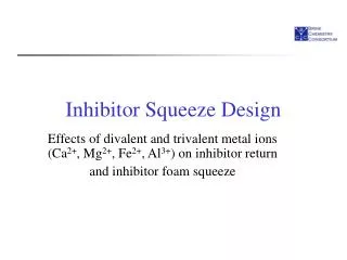
Liver ischemia/reperfusion injury and PDE5 inhibitor role Table 2: Effect of Tadalafil on some inflammatory cytokines in liver tissue homogenate. Sham N=12 Tadalafil N=12 I/R N=12 Tadalafil+I/R N=12 Groups 1341.08±80.20 1P= 0. 838 2P= 0.000 1608.00±.86.76 1P= 0.000 2P= 0.000 TNF-α Pg/gm liver tissue 1943.25±2.38 1P= 0.000 1330.25±103.33 7.50±.0.15 1P= 0.645 2P= 0.000 11.50±0.52 1P= 0.000 2P= 0.000 IL–6 Pg/gm liver tissue 28.75±2.38 1P= 0.000 7.25±0.87 9.65±0.44 1P= 0.985 2P= 0.000 16.25±2.80 1P= 0.000 2P= 0.000 ICAM-1 Pg/gm liver tissue 39.75±0.2.01 1P= 0.000 9.75±0.45 Values are Mean± SD: standard deviation. Comparison between groups done by ANOVA followed by LSD post hoc test. 1P: compared to Sham group. 2P: compared to I/R (group subjected to ischemia reperfusion injury). TNF-α (Tumor Necrosis Factor alpha), IL-6 (Interleuin-6), ICAM-1 (Intracellular adhesion molecule-1). Pg/gm liver tissue (Picogram/gram liver Tissue). Table 3: Effect of Tadalafil on some oxidative stress markers in liver tissue homogenate. Sham N=12 Tadalafil N=12 I/R N=12 Tadalafil +I/R N=12 Groups Malondialdhyde (MDA) (Nano mole /mg liver tissue) 164.25±38.93 1P= 0.744 2P= 0.000 215.25±40.35 1P= 0.018 2P= 0.000 159.25±44.88 494.25±85.72 1P= 0.000 Reduced Glutathione (GSH) (Micromole/gm liver tissue) 19.92±3.18 1P= 0. 828 2P= 0.000 15.50 ±3.50 1P= 0.003 2P= 0.000 9.00±1.28 1P= 0.000 19.50±1.3.80 Values are Mean± SD: standard deviation. Comparison between groups done by ANOVA followed by LSD post hoc test. 1P: compared to Sham group. 2P: compared to I/R (group subjected to ischemia reperfusion injury). TNF-α (Tumor Necrosis Factor alpha), IL-6 (Interleuin-6), ICAM-1 (Intracellular adhesion molecule-1). Pg/gm liver tissue (Picogram/gram liver Tissue). Table 4: Histopathological findings in the different study groups. Sham N=12 Tadalafil N=12 I/R N=12 Tadalafil + I/R N=12 Congestion -- -- + -- Hepatocyte vacuolization and necrosis -- -- ++ + Sinusoidal dilatation and congestion -- -- +++ ++ Central vein dilatation -- -- +++ ++ loss of the glycogen deposition in hepatocytes +++ +++ + ++ Effect of Tadalafil on TNF alpha, IL-6 and ICAM -1 levels in liver tissue homogenate TNF alpha, IL-6 and ICAM-1 Tadalafil achieved significant decrease in the level of TNF alpha, IL-6 and ICAM-1 in (Icariin +I/ R) when compared with IR group (Table 2). Effect of Tadalafil on some oxidative stress markers in liver tissue homogenate Tadalafil ameliorated oxidative stress by signifi- cant reduction of lipid peroxidation, as indicated by decrease of MDA levels, and restoring reduced glutathione levels but still significantly different from sham group (Table 3). Histological Assessment Tadalafil + I/R group showed majority of the he- patocytes and blood sinusoids appear preserved (Fig. 1, Table 4). Also, it showed noticeable incre- ment in glycogen content in hepatocytes (Fig. 2, Table 4). In addition, it showed decreased collagen fibers around the portal areas as well as in the pe- risinusoidal region (Fig. 3, Table 5). Immunohistochemical Assessment I/R showed negative BCL-2 expression, while Tadalafil + I/R showed positive BCL-2 expression and area percentage of BCL-2 reactions was signi- ficantly increased in Tadalafil + I/R group (Fig. 4, Table 5). I/R group showed strong positive im- munoreaction for TNF-α in the hepatocytes cyto- plasm and in the sinusoids wall. Tadalafil + I/R group showed weak immunoreaction for TNF- α in in some hepatocytes cytoplasm and in the sinu- soids wall. Optical density (OD) of TNF-α supports the descriptive findings (Fig. 5, Table 5). DISCUSSION I/R is one of the clear components of liver injury 328
H. N. Mustafa et al. Fig 1.A: Sham group showed polyhedral hepatocytes radiating from the central vein (CV), with rounded ve- sicular nuclei and acidophilic cytoplasm separated by blood sinusoids (arrow). B: Tadalafil group showed architecture being nearly similar to sham group. C: I/R group showed majority of hepatocytes around the cen- tral vein (CV) appear necrotic while the remaining ap- peared vacuolated or with acidophilic cytoplasm and dark nuclei. Also, disorganized hepatic architecture around the central vein (CV). The central veins and the blood sinusoids (arrow) are dilated and congested. D: Tadalafil + I/R group showed most of the hepatocytes and blood sinusoids (arrow) appear preserved. Some hepatocytes appeared vacuolated or with acidophilic cytoplasm and dark nuclei. The central vein (CV) and some blood sinusoids are still dilated and congested (H&E, Scale bar: 20 µm). Fig 2.A: Sham group showed positive PAS reaction of magenta staining in which glycogen is present within hepatocytes. B: Tadalafil group showed nearly similar to sham group. C: I/R group showed decreased glyco- gen storage in hepatocytes. D: Tadalafil + I/R group showed marked increment in glycogen content in hepatocytes (PAS, Scale bar: 20 µm). Fig 4.A: Sham group showed positive BCL-2 expres- sion (arrow). B: Tadalafil group showed positive BCL-2 expression (arrow). C: I/R group showed minimal BCL- 2 expression (arrow). D: Tadalafil + I/R group showed decreased collagen deposition in the portal tract area (arrow) (BCL-2, Scale bar: 20 µm). early phase is due to ischemia caused by lack of oxygen, the late phase due to reperfusion and it is characterized by the activation of Kupffer cells and the release of various mediators (Peralta et al., 2013). The current study highlights the mechanism of tadalafil toward the attenuation of liver ischemia. Serum ALT and AST levels are widely used as markers of liver cell damage. In this, tadalafil at a dose of 10 mg/kg ameliorated hepatic I/R injury, as demonstrated by reduction in AST and ALT levels, ameliorated oxidative stress status and cytokines’ profile, in addition to decreased histopathological alterations. These results are in conformity with other studies that show the beneficial effects of other phosphodiesterase inhibitors in depleting elevated ALT and AST in I/R induced liver injury Fig 3.A: Sham group showed minimal collagen around the central vein (arrow). B: Tadalafil group showed minimal collagen fibers in the portal tract area (arrow). C: I/R group showed increased deposition of collagen fibers in the portal tract area (arrow) and in the peris- inusoidal spaces. D: Tadalafil + I/R group showed de- creased collagen deposition in the portal tract area (arrow) and in the perisinusoidal spaces (Masson tri- chrome, Scale bar: 20 µm). that is evidenced in liver transplantation and liver resection. Other conditions in which I/R occur is sepsis, hepatic artery ligation, trauma and hemorr- hagic shock (Liu et al., 2016). Liver I/R injury is elicited by more than one mechanism, mainly the oxidative stress that results in damage in various organs. Injury in liver I/R consists of 2 phases. The 329
Liver ischemia/reperfusion injury and PDE5 inhibitor role Table 5: Means±SD of the area percent of collagen and BCL-2 in the studied groups. Sham N=12 Tadalafil N=12 I/R N=12 Tadalafil + I/R N=12 1.21 ± 0.6 1P<0.01 2P<0.01 3P<0.001 3.81± 0.98 1P<0.001 2P<0.001 Area percent of collagen 0.28 ± 0.04 0.31± 0.02 3.64 ± 0.04 1P<0.001 2P<0.001 3P<0.001 2.32 ± 0.08 1P<0.001 2P<0.001 Area percentage of BCL -2 4.72 ± 0.05 4.76 ± 0.02 2.51±0.12 1P<0.001 2P<0.001 3P<0.001 6.22±0.41 1P<0.001 2P<0.001 Optical density (OD) of TNF-α 0.84±0.03 0.95±0.01 1P comparison with sham, 2P comparison with Tadalafil, 3P comparison with I/R group. (Genoves et al., 2014). Cyclic nucleotides (cAMP and cGMP) are playing a pivotal role in signal transduction in many phy- siological processes, as they are working as se- cond messengers. Their Intracellular levels are controlled by adenylyl and guanylyl cyclases, which are used for their synthesis while they are degraded by PDEs (Gulati and Singh, 2014). Reactive oxygen species (ROS) are considered one of the main constituents involved in I/R. The Cellular antioxidant enzymatic and non-enzymatic defense system plays a main role in the mitigation of tissue injury elicited by free radicals. Hepatic cellular injury occurs because of the ROS's direct effect on biological components (Sehitoglu et al., 2015). Eradication of ROS in healthy cells is main- tained by a protective scavenging system that eli- minate the excessively released ROS as (CAT), superoxide dismutase (SOD), and GSH. Oxidative stress happens due to the excessive release of ROS and the decreases antioxidant defense sys- tem (Sheweita et al., 2015). GSH is oxidized with the enzymatic effect of glu- tathione peroxidase to remove the ROS, and hen- ce release oxidized glutathione (GSSG) in the he- patic cells, which explains the important role of GSH in protection against oxidative stress (Peralta et al., 2013). During I/R, GSH decreases, as the release of ROS consumes it, leading to further oxidation and degradation of vital structures in the cell as lipids, proteins and DNA (Hegazy et al., 2018). In this study, tadalafil significantly elevated the depleted GSH level attenuating the oxidative stress elicited by I/R in rats. These results are in harmony with previous studies that stated that PDE inhibitors restoring GSH level (Luo et al., 2015). Lipid peroxi- dation is another main mediator in the oxidative stress produced in different organ injuries. Oxida- tion of the lipids in cellular membranes lead to ce- llular damage and the end-product in MDA (Mustafa et al., 2015). In the present study, I/R injury resulted in an excessive amount of MDA levels in the liver. The liver MDA level was more Fig 5.A: Sham group showed negative immunoreac- tion for TNF-α protein expression in the cytoplasm of hepatocytes (arrow). B: Tadalafil group showed mini- mal immunoreaction for TNF-α (arrow). C: I/R group showed strong positive immunoreaction for TNF-α in the wall of blood sinusoids and in the cytoplasm of he- patocytes (arrow). D: Tadalafil + I/R group showed weak immunoreaction for TNF- α in the wall of blood sinusoids and in the cytoplasm of some hepatocytes (arrow) (TNF-α, Scale bar: 20 µm). than that of the normal liver, which is in harmony with other previous studies that showed the signifi- cant elevation of MDA in ischemic liver (Cakir et al., 2016). Tadalafil significantly depleted the elevated MDA levels in the liver of I/R rats, and attenuated the lipid peroxidation effectively. From the main sour- ces of ROS in I/R hepatic injury is the kupffer cells and the polymorphonuclear neutrophils (Datta et al., 2013), as their number and activity determines the severity of oxidative activity. Moreover, the activated Kupffer cells produce cytokines, espe- cially TNF-α and IL-1β (Peralta et al., 2013). In the current study, tadalafil is associated with some sinusoidal dilatation. This can be attributed to the fact that this drug is metabolized in the liver via the cytochrome P450 system (CYP 3A4 and 2C9), and a toxic or immunogenic intermediate may account for the rare instances of hepatic inju- 330
H. N. Mustafa et al. Sildenafil-associated hepatoxicity: a review of the lite- rature. Eur Rev Med Pharmacol Sci, 21: 17-22. ry (Graziano et al., 2017; Osayame and Ewek, 2011). In support of this, previous studies found that the tadalafil may be associated with some cy- toarchitectural distortion, occasional central vein and portal vessels dilatation (Osayame and Ewek, 2011; Nna et al., 2015; Jarrar and Almansour, 2015). Local and systemic inflammatory response elici- ted by I/R is controlled mainly by released cytoki- nes such as TNF-α, and affect significantly organ injury (Rao et al., 2013). During hepatic I/R, leu- kocyte recruitment is visible in liver injury, and ele- vated expression of ICAM-1 in endothelial cells promotes leukocyte adhesion and induces clot formation that changes sinusoidal perfusion (Camara-Lemarroy et al., 2014). Beside the in- creased local expression of ICAM-1 after hepatic I/ R, increased expression of ICAM-1 in other distant organs has been shown, and that explains the multiorgan failure detected after I/R (Camara- Lemarroy et al., 2014; Rao et al., 2013). Conclusion Tadalafil effects suggest that modulation of the inflammatory response might be one of the me- chanisms of tadalafil-mediated hepatoprotection. REFERENCES BEKTAS S, KARAKAYA K, CAN M, BAHADIR B, GUVEN B, ERDOGAN N, OZDAMAR SO (2016) The effects of tadalafil and pentoxifylline on apoptosis and nitric oxide synthase in liver ischemia/reperfusion inju- ry. Kaohsiung J Med Sci, 32: 339-347. GULATI P, SINGH N (2014) Tadalafil enhances the neu- roprotective effects of ischemic postconditioning in mice, probably in a nitric oxide associated manner. Can J Physiol Pharmacol, 92: 418-426. HEGAZY GA, MUSTAFA HN, ATAHI RM, YOUSEF JM (2018) The ameliorative potential of dexmedetomidine and benincasa cerifera extract in renal ischemia/ reperfusion injury in a streptozotocin-induced diabetic model. Biomed Pharmacol J, 11: 285-303. HUEPER K, LANG H, HARTLEBEN B, GUTBERLET M, DERLIN T, GETZIN T, CHEN R, ABOU-REBYEH H, LEHNER F, MEIER M, HALLER H, WACKER F, RONG S, GUELER F (2018) Assessment of liver ischemia reperfusion injury in mice using hepatic T2 mapping: Comparison with histopathology. J Magn Reson Imaging, 48: 1586-1594. JARRAR BM, ALMANSOUR MI (2015) Hepatic histolo- gical alterations and biochemical changes induced by sildenafil overdoses. Pak J Pharm Sci, 28: 2119-2127. KORKMAZ-ICOZ S, RADOVITS T, SZABO G (2018) Targeting phosphodiesterase 5 as a therapeutic option against myocardial ischaemia/reperfusion injury and for treating heart failure. Br J Pharmacol, 175: 223- 231. KUCUK A, YUCEL M, ERKASAP N, TOSUN M, KOKEN T, OZKURT M, ERKASAP S (2012) The effects of PDE5 inhibitory drugs on renal ischemia/reperfusion injury in rats. Mol Biol Rep, 39: 9775-9782. LIU A, HUANG L, GUO E, LI R, YANG J, LI A, YANG Y, LIU S, HU J, JIANG X, DIRSCH O, DAHMEN U, SUN J (2016) Baicalein pretreatment reduces liver ische- mia/reperfusion injury via induction of autophagy in rats. Sci Rep, 6: 25042. CAKIR T, ASLANER A, TEKELI SO, GÜNEŞ K, KINACI E, DOĞAN U, TEKELI F, AKYÜZ C, KOÇ S, YILMAZ N (2016) Grape seed protects cholestatic rats liver from ischemia/reperfusion injury. Acta Cir Bras, 31: 183-189. LUO M, DONG L, LI J, WANG Y, SHANG B (2015) Pro- tective effects of pentoxifylline on acute liver injury induced by thioacetamide in rats. Int J Clin Exp Pathol, 8: 8990-8996. CAMARA-LEMARROY CR, GUZMAN-DE LA GARZA FJ, ALARCON-GALVAN G, CORDERO-PÉREZ P, MUÑOZ-ESPINOSA L, TORRES-GONZÁLEZ L, FER- NÁNDEZ-GARZA NE (2014) Hepatic ischemia/ reperfusion injury is diminished by atorvastatin in Wis- tar rats. Arch Med Res, 45: 210-216. MUSTAFA HN (2016) The role of curcumin in strepto- zotocin-induced hepatic damage and the trans- differentiation of hepatic stellate cells. Tissue Cell, 48: 81-88. MUSTAFA HN, EL AWDAN SA, HEGAZY GA, ABDEL JALEEL GA (2015) Prophylactic role of coenzyme Q10 and Cynara scolymus L on doxorubicin-induced toxi- city in rats: Biochemical and immunohistochemical study. Indian J Pharmacol, 47: 649-656. DATTA G, FULLER BJ, DAVIDSON BR (2013) Molecu- lar mechanisms of liver ischemia reperfusion injury: insights from transgenic knockout models. World J Gastroenterol, 19: 1683-1698. MUSTAFA HN, HEGAZY GA, AWDAN SAE, ABDELBA- SET M (2017) Protective role of CoQ10 or L-carnitine on the integrity of the myocardium in doxorubicin indu- ced toxicity. Tissue Cell, 49: 410-426. FREITAS SH, DORIA RGS, BUENO RS, ROCHA WB, FILHO JRE, MORAES JRE, VIDANE AS, AMBRÓSIO CE (2017) Evaluation of potential changes in liver and lung tissue of rats in an ischemia-reperfusion injury model (modified pringle maneuver). PLoS One, 12: e0178665. NNA V, AKPAN U, OKON V, ATANGWHO I (2015) He- patotoxicity following separate administration of two phosphodiesterase-5 inhibitors (sildenafil and tadalafil) and opioid (tramadol); evaluation of possible reversal following their withdrawal. J Appl Pharm Sci, 5: 105- 113. GENOVES P, GARCIA D, CEJALVO D, MARTIN A, ZARAGOZA C, TOLEDO AH, TOLEDO-PEREYRA LH, LLORIS-CARSI JM (2014) Pentoxifylline in liver ischemia and reperfusion. J Invest Surg, 27: 114-124. OSAYAME A, EWEK A (2011) The effects of sildenafil citrate on the liver and kidneys of adult wistar rats (Rattus norvegicus) – A histological study. In: Sexual GRAZIANO S, MONTANA A, ZAAMI S, ROTOLO MC, MINUTILLO A, BUSARDÒ FP, MARINELLI E (2017) 331
Liver ischemia/reperfusion injury and PDE5 inhibitor role Dysfunctions - (Special Issues). IntechOpen. PERALTA SANCHO J (2013) Hepatic ischemia and reperfusion injury: effects on the liver sinusoidal milieu. J Hepatol, 59: 1094-1106. C, JIMENEZ-CASTRO MB, GRACIA- RAO J, QIAN X, WANG P, PU L, ZHAI Y, WANG X, ZHANG F, LU L (2013) All-trans retinoic acid precondi- tioning protects against liver ischemia/reperfusion inju- ry by inhibiting the nuclear factor kappa B signaling pathway. J Surg Res, 180: e99-e106. REFFELMANN T, KLONER RA (2009) Phosphodieste- rase 5 inhibitors: are they cardioprotective? Cardio- vasc Res, 83: 204-212. SAHIN T, BEGEC Z, TOPRAK HI, POLAT A, VARDI N, YÜCEL A, DURMUŞ M, ERSOY MÖ (2013) The ef- fects of dexmedetomidine reperfusion injury in rats. J Surg Res, 183: 385-390. on liver ischemia- SAVVANIS S, NASTOS C, TASOULIS MK, PAPOUT- SIDAKIS N, DEMONAKOU M, KARMANIOLOU I, AR- KADOPOULOS N, SMYRNIOTIS V, THEODORAKI K (2014) Sildenafil attenuates hepatocellular injury after liver ischemia reperfusion in rats: a preliminary study. Oxid Med Cell Longev, 2014: 161942. SAWAMURA F, KATO M, FUJITA K, NAKAZAWA T, BEARDSWORTH A (2009) Tadalafil, a long-acting inhibitor of PDE5, improves pulmonary hemodynamics and survival rate of monocrotaline-induced pulmonary artery hypertension in rats. J Pharmacol Sci, 111: 235- 243. SEHITOGLU MH, HAN H, KALIN P, GULCIN I, OZKAN A, ABOUL-ENEIN HY (2015) Pistachio (Pistacia vera L.) gum: a potent inhibitor of reactive oxygen species. J Enzyme Inhib Med Chem, 30: 264-269. SHEWEITA S, SALAMA B, HASSAN M (2015) Erectile dysfunction drugs and oxidative stress in the liver of male rats. Toxicol Rep, 2: 933-938. TAS HEKIMOGLU A, TOPRAK G, AKKOC H, EVLIYAOGLU O, OZEKINCI S, KELLE I (2013) Oxyto- cin ameliorates remote liver injury induced by renal ischemia-reperfusion in rats. Korean J Physiol Phar- macol, 17: 169-173. YE Z, CHEN O, ZHANG R, NAKAO A, FAN D, ZHANG T, GU Z, TAO H, SUN X (2015) Methane attenuates hepatic ischemia/reperfusion injury in rats through antiapoptotic, anti-inflammatory, and antioxidative ac- tions. Shock, 44: 181-187. ZHAI Y, BUSUTTIL RW, KUPIEC-WEGLINSKI JW (2011) Liver ischemia and reperfusion injury: new in- sights into mechanisms of innate-adaptive immune- mediated tissue inflammation. Am J Transplant, 11: 1563-1569. 332


![Read ebook [PDF] Fatty liver cookbook: 87 effective fatty liver diet recipes plus guide to](https://cdn7.slideserve.com/12580531/slide1-dt.jpg)