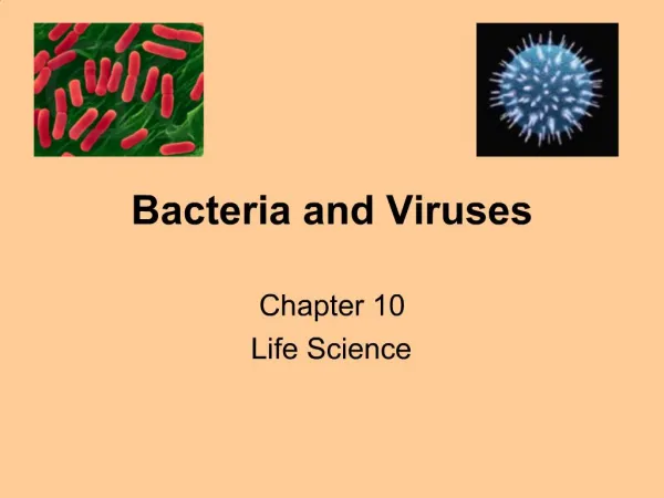
Ch. 19 Bacteria and Viruses
870 likes | 1.14k Vues
Ch. 19 Bacteria and Viruses. Ch. 19 Outline. 19-1: Bacteria Classifying prokaryotes Identifying Prokaryotes Metabolic Diversity Growth and Reproduction Importance of Bacteria. Bacteria. Once microscopes were invented, scientists discovered a world of microorganisms.

Ch. 19 Bacteria and Viruses
E N D
Presentation Transcript
Ch. 19 Outline • 19-1: Bacteria • Classifying prokaryotes • Identifying Prokaryotes • Metabolic Diversity • Growth and Reproduction • Importance of Bacteria
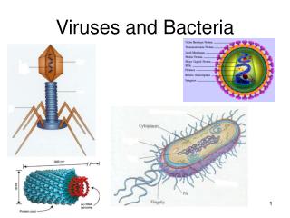
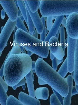
Bacteria • Once microscopes were invented, scientists discovered a world of microorganisms. • The smallest and most common microorganisms are the prokaryotes, which are single-celled organisms that lack a nucleus • The word bacteria is so familiar, that we will use it as a common term to describe prokaryotes
Ch. 19 Outline • 19-2: Viruses • What is a virus? • Viral Infection • Retroviruses • Viruses and Living Cells • 19-3 Diseases caused by bacteria and viruses • Bacterial Diseases in Humans • Controlling Bacteria • Viral Diseases • Viroids and Prions
Bacteria • Prokaryotes range in size from 1 to 5 micrometers in diameter. There are exceptions to this rule. The Epulopiscium fisheloni is about 500 micrometers long.
Classifying Prokaryotes • All prokaryotes were once classified in a single Kingdom named Monera. • Two Kingdoms of bacteria: Eubacteria, Archaebacteria • Some scientists think that the Eubacteria and Archaebacteria should be classified as Domains.
Classifying Prokaryotes • Eubacteria: the larger of the two kingdoms • Some live in fresh water, salt water, land, on and within the human body. They can infect large animals • Eubacteria cell walls protects the cell from injury and determines its shape • Eubacteria cell walls contain peptidoglycan, a carbohydrate • Inside the cell wall, a cell membrane protects the cytoplasm of eubacteria. Some eubacteria have a second cell membrane outside the cell membrane
Classifying Prokaryotes • Archaebacteria are similar to Eubacteria in that they are equally small, lack nuclei, and have cell walls • Archaebacteria do not have peptidoglycan in their cell walls, plus they have different membrane lipids • Also, the DNA sequences of key archaebacteria are more like those of eukaryotes than those of eubacteria • ****archaebacteria may possibly be ancestors of eukaryotes****
Classifying Prokaryotes • Many archaebacteria live in harsh environments. One group of arcaheabacteria is the methanogens,that produce methane gas. • Methanogens Use only CO2, H and N to produce energy to live, and as a result give off methane gas. • Live in swamps, marshes, gut of cattle, termites, etc. • Methanogens are decomposers • Other archaebacteria live in extremely salty environments or in hot springs, mud or digestive tracts of animals www.biology.iupui.edu/.../N100/2k23domain.html
Classifying Prokaryotes Bacteria are classified into the kingdoms of include a variety of lifestyles such as live in harsh environments such as
Eubacteria Archaebacteria Living in soil Infectinglarge organisms Thick mud Animal digestive tracts Salty lakes Hot springs Classifying Prokaryotes Bacteria are classified into the kingdoms of include a variety of lifestyles such as live in harsh environments such as
Identifying Prokaryotes: Shapes • Prokaryotes are identified by: • Their shape • The chemical nature of cell walls • The way they move
Identifying Prokaryotes: Shapes Rod-shaped prokaryotes are called bacilli Spherical shaped prokaryotes are called cocci Spiral and corked-shaped prokaryotes are called spirilla
Identifying Prokaryotes: Cell Walls • Two different types of cell walls are found in Eubacteria • A method called Gram stainingis used to tell the two different types of cell walls apart • Violet stain is used to stain the peptidoglycan cell walls • Alcohol treatment may wash away the violet stain. If the violet stain remains, then the bacteria are said to be Gram-positive • These bacteria have thick peptidoglycan cell walls
Identifying Prokaryotes: Cell Walls • Gram-negative bacteria have much thinner cell walls inside an outer lipid layer • Alcohol dissolves the lipid and removes the violet stain from the cell walls. The counterstain makes these bacteria appear pink
Identifying Prokaryotes: Cell Walls • What does the type of cell wall have to do with a bacterium’s resistance to antibiotics? • Gram negative bacteria’s extra layer outside the cell wall can make it hard for some antibiotics to get inside the cell (where they can work). • That makes it important for a doctor to know what kind of bacteria is causing the infection so that most effective antibiotic can be used to treat it.
Identifying Prokaryotes: Movement • How are the ways prokaryotes move? • Some do not move • Propelled by flagella • Lash snake or spiral forward • Glide • Which characteristic of prokaryotes illustrates their diversity best? By the way they obtain energy
Identifying Prokaryotes: Metabolic Diversity • Which characteristic of prokaryotes illustrates their diversity best? • By the way they obtain energy • Most prokaryotes are heterotrophs, meaning they get their energy by consuming organic molecules made by organisms • Other prokaryotes are autotrophs and make their own food from inorganic molecules
Identifying Prokaryotes: Metabolic Diversity • Most heterotrophic prokaryotes must take in organic molecules for both energy and a source of carbon. These are called chemoheterotrophs. • Humans are also chemoheterotrophs • If human food is not handled carefully, bacteria may eat our food and release toxins that cause food poisoning
Identifying Prokaryotes: Metabolic Diversity • Photoautotrophs use light energy to convert carbon dioxide and water to carbon compounds and oxygen • Photoautotrophs are found where light is plentiful • The photoautrophs Cyanobacteria contain a bluish pigment and chlorophyll. They are found everywhere (land, salt and fresh water) and are the first to recolonize an area after a natural disaster.
Identifying Prokaryotes: Metabolic Diversity • Chemoautotrophs perform chemosynthesis. • They make organic carbon molecules from carbon dioxide.
Identifying Prokaryotes: Metabolic Diversity Chemoautotrophs do not require light as a source of energy. Instead, they use energy directly from chemical reactions involving ammonia, hydrogen sulfide, nitrites, sulfur or iron Some live deep in the darkness of the ocean They use hydrogen sulfide gas that flows from hydrothermal vents in the ocean
Identifying Prokaryotes: Metabolic Diversity • Like all organisms, bacteria need a constant supply of energy. This energy is released by the process of cellular respiration or photosynthesis or both • Organisms that require a constant supply of oxygen to live are called Obligate Aerobes • Obligate AnerobesDO NOT REQUIREoxygenand may be killed by it
Clostridium botulinum Obligate Anerobe found in soil Gram positive Rod-shaped Grow in can foods that have not been properly sterilized
FaculativeAnerobes can live with or without oxygen • Faculative anerobes can live anywhere because they can switch between the processes of cellular respiration or fermentation depending on their environment
Escherichia Coli (E. Coli) • Faculative anerobe • Gram negative • Rod shaped • Lives anerobicallyin large intestine • Livesaerobicallyin sewage or contaminated water
Escherichia Coli (E. Coli) • Eschericha coli are normal inhabitants of our digestive tract • A new strain of E. coli(O157:H7) has caused illness and death for people who ate contaminated hamburger meat.
Growth and Reproduction • Bacteria can not grow and divide indefinitely because of the availability of food and they have to get rid of wastes • Bacteria grow and divide very rapidly. Their method of division is called binary fission • Bacteria grow until they double in size, copies DNA and simply splits into two daughter cells • Binary fission is just asexual reproduction (no exchange of genetic material)
Binary Fission http://www.swt.edu/~rr33/
Growth and Reproduction • Conjugation: A process of exchanging genetic info in bacteria • A hollow bridge forms between two bacterial cells and genes move from one cell to the other • Increases genetic diversity of bacteria
Growth and Reproduction • When growth conditions become unfavorable, many bacteria produce spores, which can remain dormant until there are more favorable growth conditions. • Endospore: one type of spore formed when a bacterium produces a thick internal wall that encloses its DNA and a portion of its cytoplasm.
Importance of Bacteria • We could not survive without bacteria. Some are producers, others are decomposers, and some are used by humans for various things.
Importance of Bacteria We could not survive without bacteria. Some are producers, others are decomposers, and some are used by humans for various things. Decomposers Bacteria help recycle nutrients in the environment Attack and digest dead tissue Break down complex compounds in sewage to simpler ones Produce purified water Produce nitrogen and carbon dioxide gases Produce fertilizer compounds
Importance of Bacteria Nitrogen Fixers PLANTS NEED NITROGEN TO MAKE AMINO ACIDS which are used to make PROTEINS Plants can not use nitrogen gas (N2) directly. Nitrogen must first be changed chemically to ammonia (NH3) or other nitrogen compounds. The process of converting nitrogen gas to a form that plants can use is called nitrogen fixation. Many plants have symbiotic relationships with nitrogen-fixing bacteria. The bacterium Rhizobium, grows on the roots of soybean plants. The plant provides nutrients for Rhizobium, and it converts nitrogen gas in the air to ammonia, which helps the plant.
Importance of Bacteria • Human Uses of Bacteria • Produce a wide variety of food and beverages • Industry cleaning up oil spills (digest petroleum) • Mine minerals from the ground • Remove wastes and poisons from water • Synthesize drugs and chemicals (genetic engineering) • Produce vitamins in human intestines • Produce heat stable enzymes that can be used in medicine, food production, and industrial chemistry
Ch. 19 Outline • 19-2: Viruses • What is a virus? • Viral Infection • Retroviruses • Viruses and Living Cells
What is a Virus? • The word virus is derived from the Latin word forpoison • Dmitri Ivanovski identified the cause of tobacco mosaic disease by extracting juice from an infected plant • Martinus Beijerinck suggested that tiny particles in the extracted juicecaused tobacco mosaic disease (called particles viruses) • Wendell Stanley inferred that viruses were not alive when he obtained crystals of tobacco mosaic virus
What is a Virus? The word virus is derived from the Latin word forpoison Viruses: particles of nucleic acids, proteins and in some cases, lipids Viruses are NON-LIVING, but they do reproduce. Viruses can reproduce only by infecting living cells and once inside, they use the machinery of the infected cell to produce more viruses
Viral Structures • A typical virus is composed of a core of DNA or RNA surrounded by a protein coat.
What is a Virus? • The simplest virusmay have only a few genes, whereas the most complexmay have more than a hundred genes • A viruses protein coat is called its capsid. The capsid includes proteins that enable a virus to enter a host cell. • The capsid proteins of a typical virus bind to receptors on the surface of a cell and “trick” the cell into allowing it inside.
What is a Virus? • Once inside, the viral genes are expressed. The cell transcribes and translates the viral genetic information into viral capsid proteins • Sometimes the genetic program causes the host cell to make copies of the virus, and in the process the host cell is destroyed.
What is a Virus? Viruses must bind precisely to proteins on the cell surface and then use the host’s genetic system Viruses that infect bacteria are called bacteriophages
Viral Infection • There are two types of viral infections: lytic and lysogenic • Lytic Infection • The virus enters the cell, makes copies of itself, and causes the cell to burst (destroyed) • In a lytic infection, the protein capsid is activated by contact with a specific host cell • It then injects its DNA directly into the host cell. • The host cell cannot tell the difference between its own DNA and the DNA of the virus
Viral Infection Consequently, the cell begins to make messenger RNA from the genes of the virus This viral mRNA is translated into viral proteins that act like a molecular wrecking crew, chopping up the cell DNA, a process that shuts down the infected host cell In this lytic infection, the virus then uses the materials of the host cell to make thousands of copies of its own DNA molecule