Download
1 / 43
470 likes | 803 Vues
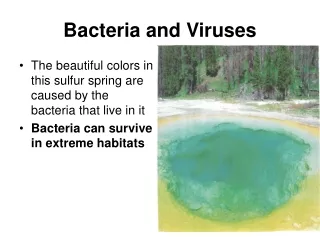
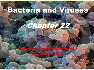
Bacteria and Viruses. Chapter 22 Mountain Pointe High School R. LeBlanc, M.S. How small is life?. 100 µm. 20 µm. 0.5 µm. What does um represent? What do the yellow structures represent and how did they get there? How large are these bacillus bacterial cells?. bacteriophage.

E N D
Bacteria and Viruses Chapter 22 Mountain Pointe High School R. LeBlanc, M.S.
How small is life? 100 µm 20 µm 0.5 µm • What does um represent? • What do the yellow structures represent and how did they get there? • How large are these bacillus bacterial cells?
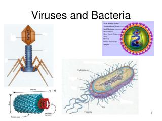
bacteriophage ruptured bacterial cell • Can you see the bacteria? • What are bacteriophage? • Which are larger, virus or bacteria? How much smaller? • Are these viruses living? Why or why not? Fig. 22.2, p. 355 1.5 µm
Metabolic Diversity Photoautotrophic (Energy from: CO2; H2O; H2; H2S) Chemoautotrophic(Energy from: CO2; H2; S; NH4) Photoheterotrophs (carbon source=fatty acids; carbs) Chemoheterotrophic(parasites or saprobes) Sizes and Shapes (all of these are unicellular) 1 - 10 micrometers Coccus Bacillus Spirillum Characteristics of Bacteria What is the difference between an autotroph and heterotroph?
coccus bacillus Small staffs maybe skiny or fat Sometimes oval & flattened spirillum One or more twist; some like a comma; flexible or stiff corkscrews in-text, p. 356
Structures Cell walls Peptidoglycan (molecules of polysaccharide cross linked with short polypeptides) Membrane (some membrane compartments: plasma) Characteristics of Bacteria
Characteristics of Bacteria • Glycocalyx (mesh that encloses wall; called capsule) • DNA in cytoplasm (not membrane bound) • Flagella (movement; one or more; whip like) • Gram stain (use to ID bacteria: refer to other slide)
0.5 µm 1 µm • H. pylori with its many flagella • Pathogenic bacteria • Found in: water, food, esp. unpasteurized milk. • Bacilli & coccus bacteria attached to human teeth. (Are these bacteria harmful?) • E. coli dividing; Pili-filament projections Fig. 22.4, p. 357 0.5 µm
Bacterial Structure Pili are protein filaments that help bacteria to adhere to surfaces; aids in conjugation (sexual reproduction)
Proper Gram Staining Procedures: • Collect bacterial sample and carefully heat-fix cells to microscope slide. • Add Crystal Violet, a purple dye, to the bacterial sample turning all cells purple. • Then add iodine, a binding agent, that causes the purple dye to stick to the gram positive bacterial cells. • Use an alcohol wash to rinse off the purple dye from the gram negative cells making them neutral in color. • Last, counter-stain the bacterial sample with safranin, a pink dye, that will stick to the gram negative cells turning them pink. • Gram Positive: bacteria cells purple. • Gram Negative: bacteria cells pink with counter stain. stain with purple dye stain with iodine wash with alcohol counterstain with safranin
Prokaryotic Fission • When nearly doubled in size, it divides; 10-30 minutes. • In some cases budding takes place; daughter cell buds off parent cell • NOTICE: Cell synthesizes protein & lipid molecules causing the plasma membrane to grow moving the 2 DNA molecules apart. • Original DNA is a single circular chromosome thread.
a Bacterium (cutaway view) before DNA replication. The bacterial chromosome is attached to the plasma membrane. b DNA replication starts. It proceeds in two directions away from the same site in the bacterial chromosome. c The new copy of DNA is attached at a membrane site near the attachment site of the parent DNA molecule. d New membrane grows between the two attachment sites. As it increases, it moves the two DNA molecules apart. e At the cell midsection, deposits of new membrane and new wall material extend down into the cytoplasm. f The ongoing, organized deposition of membrane and wall material at the cell midsection divides the cell in two IDENTICAL DAUGHTER CELLS. Fig. 22.7, p. 358
BACTERIAL CONJUGATION nicked plasmid conjugation tube a A conjugation tube has already formed between a donor and a recipient cell. An enzyme has nicked the donor’s plasmid. b DNA replication starts on the nicked plasmid. The displaced DNA strand moves through the tube and enters the recipient cell. Plasmid: a small self-replicating circle of extra DNA & has few genes. http://www.hhmi.org/biointeractive/animations/conjugation/conj_frames.htm c In the recipient cell, replication starts on the transferred DNA. d The cells separate from each other; the plasmids circularize. GENETICALLY IDENTICAL DAUGHTER CELLS. • Conjugation: the transfer of DNA from one cell to another. • Takes place in Salmonella; streptococcus & E. coli bacteria
Bacterial Classification • Numerical taxonomy • Bacteria are not well represented in the fossil record. • Traits of unidentified bacterial cells are compared to known bacteria. • Traits include: cell shape, motility, staining attributes, nutritional requirements, metabolic patterns, endospores or not? • Gene sequencing and comparative biochemistry are used today in classifying. • Especially using rRNA • Small rRNA changes can be measured & used to relate some groups. • Newest technique to ID bacteria; nucleotide sequencing
Live in harsh conditions With distinct defined organelles eu= ‘typical’ EUBACTERIA ARCHAEBACTERIA EUKARYOTES • The definition of ‘species’ that fits sexually reproducing organisms dos NOT fit bacteria. • The term ‘strain’ is used to show minor differences between bacteria that are closely related.
Archaebacteria 1st living cells; no peptidoglycan in cell walls; found in unusual places. • Methanogens (“methane-makers”) • Swamps, sewage, mud, & animal guts. • Make ATP anaerobically: CO2 to CH4 • Halophiles (“salt-lovers”) • Brackish ponds, salt lakes, hydrothermal seafloor vents • Extreme Thermophiles (“heat-lovers”) • Acidic soils, hot springs, coal mines, hydrothermal vents
Methano-coccus • Heat-loving and methane producer Methanogen with thick polysaccharide walls called peptidoglycan.
Which major archaebacteria group is represented below???? Great Salt Lake; Which bacteria live there? Cows belch producing a unique smell. Commercial seawater evaporating ponds. Hypersaline condition. Hot, sulfur-rich water in Emerald Pond, Yellowstone National Park.
Photoautotrophic Cyanobacteria (blue green algae) Ponds and freshwater (see next slide) Chemoautotrophic Environment Cycling of N2 , S2 Building blocks of amino acids (proteins) Without nitrogen there would be NO LIFE. Plants use nitrogen fixing bacteria to recycle nitrogen. Eubacteria NOTE: They have fatty acids incorporated into their plasma membrane.
Chemoheterotrophic Most bacteria fall into this category. Pseudomonads (decompose organic even pesticides Lactobacillus (‘good’ bacteria; making pickles, yogurt, buttermilk; sauerkraut) E. Coli (produce vitamin K/other compounds) Pathogenic (Disease causing; some E. Coli; Botolinum Endospores (Tetanus found in the soil) Eubacteria
Cyanobacteria • Nutrient rich pond • Heterocyst: modified cells that form when nitrogen compounds are scarce; make a nitrogen-fixing enzyme resting spore heterocyst 5 µm
developing endospore (Resting structure) 2.2 µm • Triggered by the depletion of nitrogen or other nutrients. • When plasma membrane breaks it releases many free spores. • Can remain dormant for decades. • When conditions are favorable they become active as a bacterial cell.
Facts About Bacteria • Also classified by their arrangements. Some exist alone, most are grouped together: • Diplo - paired cells • Staphylo - clustered cells • Strepto - cells in chains
What causes this condition? Rocky Mountain Spotted Fever • Moves from host to host inside the gut of insect (tick) as bacteria. • Penetrates the cytoplasm & nucleus of host cells. • Lyme Disease: transmitted from deer ticks.
Different examples of bacterial infections Match the picture with the bacterial infection: A) LEPROSY B) LYME DISEASE C) PINK EYE D) ATHELETES FEET
0.25 µm Magnetotactic Bacterium Myxobacteria with fruiting bodies
The Viruses • Non-cellular infectious agent • Infect: cats, cattle, birds, insects, plants, fungus, protist, & bacteria (can infect organisms in ALL kingdoms). • Protein coat surrounding a nucleic acids core • Rod-like or polyhedral shapes • Used for protection, shape can change, used to attach to host cells (attach to proteins in plasma membrane of host. • DNA or RNA • Reproduce inside a host cell • Enveloped or non-enveloped
viral RNA Polyhedral Virus protein subunits of coat • Tobacco Mosaic Virus • Helical Virus 18-nm diameter, 250-nm length 80-nm diameter lipid envelope; proteins span the envelope, line its inner surface, and spike out above it DNA protein coat sheath viral RNA base plate tail fiber reverse transcriptase viral coat (proteins) Fig. 22.16, p. 364 65-nm diameter head, 225-nm total length 100-120 nm diameter
Viruses • Shape • Helical (Rod Shaped) • Polyhedral • Enveloped or non-enveloped • Spiked (some) • Complex • Viruses are host specific • Bacteriophages • Used to study viruses • Reproduce rapidly Polyhedral Virus
Viruses • Bacteriophage • Infects bacteria • Used in early experiments to determine function of DNA
Viruses • Enveloped virus • Envelope is made mostly of membrane remnents from previously infected cell • HIV is example • Trigger for AIDS • Attacks certain W.B.C. • Weakens immune system
Prions (8 rare diseases of nervous system) Small Proteins Altered products of a gene found at the surface of neurons of nervous system. Mad Cow Disease (BSE)* Diseases Kuru (brain) Scrapie (sheep); named after sheep scrape off their wool. Creutzfeldt-Jakob disease (destroys muscle & brain function) Infectious Agents Tinier Than Viruses(more stripped down than viruses) *Outbreak in 1996. Caused by ground up sheep with Scrapie where feed to cattle in turn spread to cattle, then to humans.
Infectious Agents Tinier Than Viruses • Viroids • Tight folds or circles of RNA • Plant diseases • Destroy million of $ of: • Potatoes • Citrus • Other cash crops.
5 Steps Attachment Penetration Replication Assembly Release Lytic pathway Host cell lysis Lysogenic pathway Viral DNA integrates into bacterial chromosome Viral Multiplication Cycles
In Conclusion • After the origin of life, a divergence occurred leading to Eubacteria and common ancestors of Archaebacteria and Eukaryotic cells • All bacteria are prokaryotes • Bacteria have 3 basic shapes: cocci, bacilli, and spirilla
In Conclusion • Many bacteria have external structures that increase their survival and pathogenicity • Bacteria reproduce by binary fission • Many species have plasmids and some can transfer genetic information through the process of conjugation • Bacteria as a group have metabolic diversity
In Conclusion • Viruses are nonliving infectious agents • Viruses consist of either DNA or RNA surrounded by a protein coat • Some may have an envelope and spikes • Viruses cannot reproduce on their own but must use a host cell’s machinery • There are five steps in the multiplication cycle of a virus
In Conclusion • There are two pathways common in the multiplication of bacteriophages: lytic and lysogenic • Multiplication cycles of viruses are diverse; may occur rapidly or can enter a latent phase • developed by M. Roig