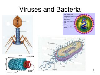
Viruses and Bacteria
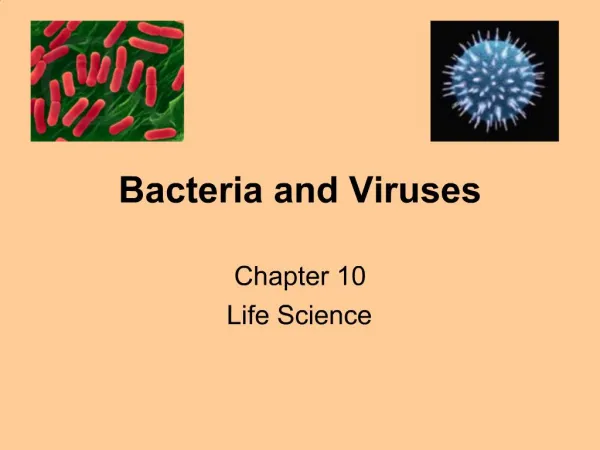
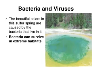
This article explores the fundamental differences between viruses and bacteria, two distinct forms of life with unique structures and reproduction methods. Bacteria are prokaryotic, single-celled organisms with peptidoglycan cell walls and a variety of shapes, including rod, spiral, and spherical. Viruses, on the other hand, are acellular entities that rely on host cells for reproduction and are much smaller than bacteria. We will discuss their definitions, structures, and the significant implications for health and medicine.

Viruses and Bacteria
E N D
Presentation Transcript
The Difference in Definition Pro karyo • Bacteria: Prokaryotic Organisms • Pro: Primitive or “prior to” • Karyon: Nucleus or “kernel” • Single-celled organisms • Has circular DNA; often has “plasmid” DNA that helps codes for genes to increase fitness (ex. Antibiotic resistance) • Viruses: Submicroscopic, parasitic, acellular entity composed of a nucleic acid core surrounded by a protein coat. • Below the resolution of a microscope • Relies on a host • Does not have the properties of cellular life Submicroscopic parasitic acellular
The Difference in Size • Bacteria can be measured in micrometers • 0.000001m or 10-6 • Viruses are measured in nanometers • 0.000000001m or 10-9
Virus Bacterium Animalcell Animal cell nucleus 0.25 m Comparing the size of a virus, a bacterium, and an animal cell
Bacteria Two main “domains” or groups • Bacteria Cell walls with peptidoglycan • Archaebacteria Cell walls lack peptidoglycan Adapted to extreme environments: - Extremely hot and cold, salty, without oxygen, etc. peptidoglycan Made up of types of peptide andsugar bonds
Bacteria: Shapes • Three basic shapes: • Rod-shaped (Bacilli) Bacillus anthracis (Anthrax),Yersinia pestis (Bubonic plague) • Comma-shaped (Vibrios) Vibrio cholerae • Spherical (Cocci) Streptococcus, Staphylococcus • Spiral (Spirilla) Treponema pallidum (Syphillis)
Bacterial Staining • Gram-positive: Retains the crystals of violet dye in the peptidoglycan layer • Infection by this type can be treated by antibiotics such as penicillin
Bacterial Staining • Gram-negative: Will not pick up the violet dye • Infection by this type must be treated by a broad-spectrum antibiotic such as ciprofloxacin peptidoglycan
Bacterial Growth and Reproduction • Binary Fission: (video) Asexual division DNA replicates and cytoplasm divides • Conjugation (video) “Sexual” reproductionSex Pilus extends between bacteria plasmid DNA is transferred from one bacterium to another • Spore Formation:occurs when growth conditions are unfavorable An endospore is a “spore” with a thick internal wall of membrane that encloses and protects its DNA
Capsomereof capsid Membranousenvelope RNA Capsomere DNA Head Capsid Tail sheath DNA RNA Tail fiber Glycoprotein Glycoprotein 80 225 nm 18 250 mm 80–200 nm (diameter) 70–90 nm (diameter) 50 nm 20 nm 50 nm 50 nm (d) Bacteriophage T4 (a) Tobacco mosaic virus (b) Adenoviruses (c) Influenza viruses Viral Shapes and structure
Viruses Reproduction Viruses reproduce by infecting other cells. Two types of viral infections: 1. Lytic Infection 2. Lysogenic Infection
0.5 m Figure 18.1 T4 bacteriophage infecting an E. coli cell
The Lytic Infection Step 1: Attachment of virus to host cell Step 5: New viruses “lyse” the host cell and are released for further infection Step 3: Replication of viral DNA and Synthesis of Protein Capsule using cellular “machinery” –DNA and RNA polymerases, ribosomes, etc. Step 4: Assembly of new viruses inside host cell Step 2: Injection of viral DNA into cell
Characteristics of Lytic Infections • Fast acting • Symptoms emerge within 24 – 48 hours • Examples – influenza, west-nile
The Lysogenic Infection Step 1: Virus attaches and inserts its DNA inside host Step 2: Viral DNA attaches to the host DNA (prophage) Step 3: The viral DNA lies “dormant” and only replicates each time the cell replicates Step 4: Stress or other “factors” causes the infection to progress to the “lytic” phase
Characteristics of Lysogenic Infections • Slow Acting - Viral DNA can lie “dormant” for many years as proghage • The host are “symptom-free” during dormancy • Infection is fast acting when the infection progresses to the lytic phase 4. Example: HIV