Download
1 / 32
320 likes | 336 Vues
This review delves into protein structures, from primary to quaternary, emphasizing structure-function relationships and computational modeling. Learn about the intricate three-dimensional analysis in laboratory settings.

E N D
CS 177 Proteins I (Structure-function relationships) Review of protein structures Computational modeling Three-dimensional structural analysis in laboratory Review of protein structures Computational Modeling Three-dimensional structural analysis in laboratory
(very) basic advanced Structure-function relationships Recommended readings A science primer: Molecular modeling http://www.ncbi.nlm.nih.gov/About/primer/molecularmod.html Brown, S.M. (2000) Bioinformatics, Eaton Publishing, pp. 99-119 Veeramalai, M. & Gilbert, D.: Bioinformatics Tools for protein structure visualisation and analysis http://www.brc.dcs.gla.ac.uk/~mallika/Publications/scwbiw-article.htm Mount, D.W. (2001) Bioinformatics,Cold Spring Harbor Lab Press, pp.382-478 Review of protein structures Computational Modeling Three-dimensional structural analysis in laboratory
Polypeptide chain The structure of two amid acids Review of protein structure Primary structure Proteins are chains of amino acids joined by peptide bonds The N-C-C sequence is repeated throughout the protein, forming the backbone Review of protein structures Computational Modeling Three-dimensional structural analysis in laboratory The bonds on each side of the C atom are free to rotate within spatial constrains,the angles of these bonds determine the conformation of the protein backbone The R side chains also play an important structural role
Review of protein structure Secondary structure: Interactions that occur between the C=O and N-H groups on amino acidsMuch of the protein core comprises helices and sheets, folded into a three-dimensional configuration:- regular patterns of H bonds are formed between neighboring amino acids- the amino acids have similar angles- the formation of these structures neutralizes the polar groups on each amino acid- the secondary structures are tightly packed in a hydrophobic environment- Each R side group has a limited volume to occupy and a limited number of interactions with other R side groups sheet helix Review of protein structures Computational Modeling Three-dimensional structural analysis in laboratory
Secondary structure Other Secondary structure elements(no standardized classification) - random coil - loop - others (e.g. 310 helix, -hairpin, paperclip) Super-secondary structure Review of protein structures Computational Modeling Three-dimensional structural analysis in laboratory - In addition to secondary structure elements that apply to all proteins (e.g. helix, sheet) there are some simple structural motifs in some proteins - These super-secondary structures (e.g. transmembrane domains, coiled coils, helix-turn-helix, signal peptides) can give important hints about protein function
A: No, because the detailed packing of the amino acid side chains is not revealed from this information. However, the Psi and Phi angles do determine the entire secondary structure of a protein Tertiary structure Review of protein structure Q: If we have all the Psi and Phi angles in a protein, do we then have enough information to describe the 3-D structure? Review of protein structures Computational Modeling Three-dimensional structural analysis in laboratory
Tertiary structure • The tertiary structure describes the organization in three dimensionsof all the atoms in the polypeptide • The tertiary structure is determined by a combination of different types of bonding:- Ionic interactions between oppositely charged residues can pull them together, • Hydrogen Bonds - Hydrogens are partially positively charged, are attracted to partially negative oxygens. (weaker) • - van Der Waals - hydrophobic residues become attractive to each other when forced together by exclusion from the aqueous surroundings. (weakest) • Many of these bonds are very week and easy to break, but hundreds or thousands working together give the protein structure great stability • If a protein consists of only one polypeptide chain, this level then describes thecomplete structure Review of protein structures Computational Modeling Three-dimensional structural analysis in laboratory
Tertiary structure Review of protein structures Computational Modeling Three-dimensional structural analysis in laboratory
Tertiary structure Proteins can be divided into two general classes based on their tertiary structure: - Fibrous proteins have elongated structure with the polypeptide chains arranged in long strands. This class of proteins serves as major structural component of cells Examples: silk, keratin, collagen - Globular proteins have more compact, often irregular structures. This class of proteins includes most enzymes and most proteins involved in gene expression and regulation Review of protein structures Computational Modeling Three-dimensional structural analysis in laboratory
Quaternary structure The quaternarystructure defines the conformation assumed by a multimeric protein.The individual polypeptide chains that make up a multimeric protein are often referred toas protein subunits. Subunits are joined by ionic, H and hydrophobic interactions Example:Haemoglobin(4 subunits) Review of protein structures Computational Modeling Three-dimensional structural analysis in laboratory
Primary structure: Sequence of amino acids Secondary structure: Interactions that occur betweenthe C=O and N-H groups on amino acids Tertiary structure: Organization in three dimensions of all the atoms in the polypeptide Quaternary structure: Conformation assumed by a multimeric protein The four levels of protein structure are hierarchical:each level of the build process is dependent upon the one below it Summary protein structure Review of protein structures Computational Modeling Three-dimensional structural analysis in laboratory
Summary protein structure Review of protein structures Computational Modeling Three-dimensional structural analysis in laboratory
Structure displays Common displays are (among others) cartoon, spacefill, and backbone backbone spacefill cartoon Review of protein structures Computational Modeling Three-dimensional structural analysis in laboratory
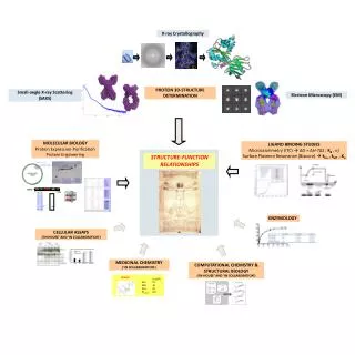
Need for analyses of protein structures A protein performs metabolic, structural, or regulatory functions in a cell. Cellular biochemistry works based on interactions between 3-D molecular structures The 3-D structure of a protein determines its function Therefore, the relationship of sequence to function is primarily concerned with understanding the 3-D folding of proteins and inferring protein functions from these 3-D structures(e.g. binding sites, catalytic activities, interactions with other molecules) The study of protein structure is not only of fundamental scientific interest in terms of understanding biochemical processes, but also produces very valuable practical benefits Medicine The understanding of enzyme function allows the design of new and improved drugs Agriculture Therapeutic proteins and drugs for veterinary purposes and for treatment of plant diseases Industry Protein engineering has potential for the synthesis of enzymes to carry out various industrial processes on a mass scale Review of protein structures Computational Modeling Three-dimensional structural analysis in laboratory
Sources of protein structure information 3-D macromolecular structures stored in databases The most important database: the Protein Data Bank (PDB)The PDB is maintained by the Research Collaboratory for Structural Bioinformatics (RCSB) and can be accessed at three different sites (plus a number of mirror sites outside the USA): - http://rcsb.rutgers.edu/pdb (Rutgers University)- http://www.rcsb.org/pdb/ (San Diego Supercomputer Center)- http://tcsb.nist.gov/pdb/ (National Institute for Standards and Technology) It is the very first “bioinformatics” database ever build Review of protein structures Computational Modeling Three-dimensional structural analysis in laboratory
Sources of protein structure information Experimental structure determination In practice, most biomolecular structures (>99% of structures in PDB) are determined using three techniques:- X-ray crystallography (low to very high resolution) Problem: requires crystals; difficult to crystallize proteins by maintaining their native conformation; not all protein can be crystallized; Review of protein structures Computational Modeling Three-dimensional structural analysis in laboratory
X-ray crystallography Review of protein structures Computational Modeling Three-dimensional structural analysis in laboratory
Sources of protein structure information Experimental structure determination In practice, most biomolecular structures (>99% of structures in PDB) are determined using three techniques:- X-ray crystallography (low to very high resolution) Problem: requires crystals; difficult to crystallize proteins by maintaining their native conformation; not all protein can be crystallized; - Nuclear magnetic resonance (NMR) spectroscopy of proteins in solution (medium to high resolution) Problem: Works only with small and medium size proteins (~50% of proteins cannot be studied with this method); requires high solubility - Electron microscopy and crystallography (low to medium resolution) Problem: (still) relatively low resolution Review of protein structures Computational Modeling Three-dimensional structural analysis in laboratory Experimental methods are still very time consuming and expensive; in most cases the experimental data will contain errors and/or are incomplete. Thus the initial model needs to be refined and rebuild
Sources of protein structure information Computational Modeling Researches have been working for decades to develop procedures for predicting protein structure that are not so time consuming and not hindered by size and solubility constrains. As protein sequences are encoded in DNA, inprinciple, it should therefore be possible to translate a gene sequence into an amino acid sequence, and topredict the three-dimensional structure of the resulting chain from this amino acid sequence Review of protein structures Computational Modeling Three-dimensional structural analysis in laboratory
If we were able to evaluate 109 conformations per second, this would still keep us busy 4 x 10259 times the current age of the universe There are optimized ab initio prediction algorithms available as well as fold recognition algorithms that use threading (compares protein folds with know fold structures from databases), but the results are still very poor Computational modeling How to predict the protein structure? Ab initio prediction of protein structure from sequence: not yet. Problem: the information contained in protein structures lies essentially in theconformational torsion angles. Even if we only assume that every amino-acid residuehas three such torsion angles, and that each of these three can only assume oneof three "ideal" values (e.g., 60, 180 and -60 degrees), this still leaves us with 27possible conformations per residue. For a typical 200-amino acid protein, this would give 27200 (roughly 1.87 x 10286)possible conformations! Q: Can’t we just generate all these conformations, calculate their energy and see which conformation has the lowest energy? Review of protein structures Computational Modeling Three-dimensional structural analysis in laboratory
Computational modeling Solution: homology modeling Homology (comparative) modeling attempts to predict structure on the strengthof a protein’s sequence similarity to another protein of known structure Basic idea: a significant alignment of the query sequence with a target sequence from PDB is evidence that the query sequence has a similar 3-D structure (current threshold ~ 40% sequence identity). Then multiple sequence alignment and pattern analysis can be used to predict the structure of the protein Review of protein structures Computational Modeling Three-dimensional structural analysis in laboratory
Computational modeling Flow chart for protein structure prediction (from Mount, 2001) Review of protein structures Computational Modeling Three-dimensional structural analysis in laboratory
Computational modeling Protein sequence - partial or full sequences; predicted through gene finding Review of protein structures Computational Modeling Three-dimensional structural analysis in laboratory
Computational modeling Database similarity search - sequence is used as a query in a database similarity search against proteins in PDB Review of protein structures Computational Modeling Three-dimensional structural analysis in laboratory
Computational modeling • Does the sequence align with a protein of known structure? • Yes: if the database similarity search reveals a significant alignment between the query sequence and a PDB target sequence, the alignment can be used to position the amino acids of the query sequence in the same approximate 3-D structure • No: proceed to protein family analysis Review of protein structures Computational Modeling Three-dimensional structural analysis in laboratory
Computational modeling • Protein family analysis/relationship to known structure • Family (structural context): structures that have a significant level of structural similarity but not necessarily significant sequence similarity • the goal is to exploit these structure sequence relationships; two questions: 1) is the new protein a member of a family, 2) does the family have a predicted structural fold? • analyze sequence for family specific profiles and patterns. Available databases: 3D-Ali, 3D-PSSM, BLOCKS, eMOTIF, INTERPRO, Pfam …) • if the family analysis reveals that the query protein is a member of a family with a predicted structural fold, multiple alignment can be used for structural modeling Review of protein structures Computational Modeling Three-dimensional structural analysis in laboratory
Computational modeling • Protein family analysis/relationship to known structure • if the family analysis is unsuccessful, proceed to structural analyses Review of protein structures Computational Modeling Three-dimensional structural analysis in laboratory
Computational modeling • Structural analysis • several different types of analyses to infer structural information • presence of small amino acid motifs in a protein can be indicator of a biochemical function associated with a particular structure. Motifs are available from the Prosite catalog • spacing and arrangement of amino acids (e.g. hydrophobic amino acids) provide important structural clues that can be used for modeling • certain amino acid combinations can occur in certain types of secondary structure • - These structural analyses can provide clues as to the presence of active sites and regions of secondary structure. These information can help to identify a new protein as a member of a known structural class Review of protein structures Computational Modeling Three-dimensional structural analysis in laboratory
Computational modeling • 3-D structural analysis in lab • proteins that fail to show any relationship to proteins of known structure are candidates for structural analyses (X-ray crystallography, NMR). There are about 600 known fold families and new structures are frequently found to have already known structural fold. Accordingly, protein families with no relatives of known structure may represent a novel fold Review of protein structures Computational Modeling Three-dimensional structural analysis in laboratory
Computational modeling: summary Partial or full sequencespredicted through gene finding Similarity searchagainst proteins in PDB Find structures that have a significantlevel of structural similarity (but notnecessarily significant sequence similarity) Alignment can be used to position theamino acids of the query sequence inthe same approximate 3-D structure If member of a family with a predicted structural fold, multiple alignment can be used for structural modeling Review of protein structures Computational Modeling Three-dimensional structural analysis in laboratory Infer structural information (e.g. presence of smallamino acid motifs; spacing and arrangement ofamino acids; certain typical amino acid combinationsassociated with certain types of secondary structure)can provide clues as to the presence of active sites andregions of secondary structure Structural analyses in the lab(X-ray crystallography, NMR)
Computational modeling: summary How to predict the protein structure? Ab initioprediction of protein structure from sequence Homology (comparative) modeling attempts to predict structure on the strength of a protein’s sequence similarity to another protein of known structure Experimental structure determination Review of protein structures Computational Modeling Three-dimensional structural analysis in laboratory
Computational modeling Viewing protein structures A number of molecular viewers are freely available and run on most computer platforms and operating systemsExamples: Cn3D 4.1 (stand-alone) Rasmol (stand-alone) Chime (Web browser based on Rasmol) Swiss 3D viewer Spdbv (stand-alone) All these viewers can use the PDB identification code or the structural file from PDB Review of protein structures Computational Modeling Three-dimensional structural analysis in laboratory