Chapter 12 Heart Cardiovascular System
Chapter 12 Heart Cardiovascular System. Overview to the circulatory system A two-way irrigation system. A pump ( heart ) cycles nutrients and oxygen out to the fields ( tissues and organs ). Depleted fluids return from the fields, dump their waste gas


Chapter 12 Heart Cardiovascular System
E N D
Presentation Transcript
Chapter 12 Heart Cardiovascular System
Overview to the circulatory system A two-way irrigation system. A pump (heart) cycles nutrients and oxygen out to the fields (tissues and organs). Depleted fluids return from the fields, dump their waste gas (CO2) in the lungs, pick up fresh oxygen, then cycle nutrients and O2 out to the fields again. The cycle repeats itself 60-80 times each minute.
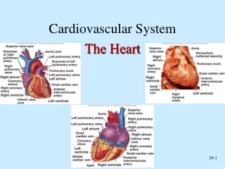
Irrigation System “Pipes” (blood vessels) Away from the heart: arteries (except Toward the heart: veins pulmonary system) arteries-- (aorta is largest) arterioles-- capillaries-- venules-- veins (vena cava is largest)
Functions of the Heart • Generate blood pressure • Assist in transport of blood • pulmonary circuit • systemic circuit • Guarantee one-way flow of blood • Regulates blood supply
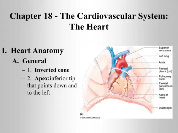
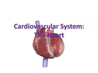
Size, Form, and Location of the Heart • Location • Thoracic cavity (mediastinum) • Must know as a health care worker! • Form: • Triangular organ • Size • ~fist-sized (5.5 x 3.5 “)
Anatomy of the Heart • Pericardium • Pericardial cavity • Pericardial fluid • Pericardium/ pericardial sac • 2 tissue layers: • Outer fibrous pericardium • Inner serous pericardium – 2 parts: • Parietal pericardium • Visceral pericardium/ epicardium
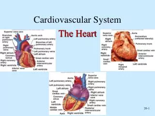
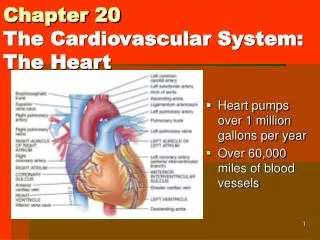
Anatomy of the Heart, continued… • External Anatomy • Atria • Ventricles • Coronary Sulcus • Superior & Inferior Vena Cava • Pulmonary Veins • Pulmonary Trunk • Pulmonary Arteries • Aorta
Anatomy of the Heart, continued… • Blood Supply • Coronary Arteries • Coronary Artery Disorders • Cardiac Veins
Anatomy of the Heart, continued… • Heart Chambers & Internal Anatomy • Right & Left Atria • Right & Left Ventricles
Anatomy of the Heart, continued… • Heart Valves • Atrioventricular valves: tricuspid, bicuspid, aortic & pulmonary semilunar • Papillary muscles & chordae tendineae • Skeleton of heart
F. Path of blood through the heart (The Voyage of the Mighty RBC) • Vena cava (superior and inferior) • 2. Right atrium • 3. Pass through right atrioventricular valve • (tricuspid valve) • 4. Right ventricle • 5. Pulmonary semi-lunar valve • 6. Pulmonary artery (O2-poor blood) • 7. Lungs (dump waste products CO2)
8. Lungs (pick up oxygen) 9. Enter left atrium via pulmonary vein (via four entrances) 10. Pass through left atrioventricular valve (bicuspid or mitral valve) 11. Left ventricle 12. Leave through aortic semilunar valve (aorta) 13. Travel throughout body, delivering O2 and nutrients, gathering waste produces such as CO2
Histology of the Heart • Heart Wall • Epicardium • Myocardium • Endocardium • Cardiac Muscle • Branched • Striated • ATP • Intercalated disks • Coordinated contractions
Action Potentials Depolarization Early repolarization & plateau phases Repolarization Refractory period Cardiac muscle fibers can contract rhythmically on their own, but must be coordinated by electrical signals (impulses) for effective pumping. (orchestra conductor) Heart has its own built-in conduction system for initiating (“starting”) and conducting (“sending) impulses through the myocardium. SA node to AV node (slow travel); AV bundle to Purkinje fibers is rapid travel Autonomic nerve signals: control heart rate Electrical Activity of the Heart
Conduction System of the Heart S-A (sino-atrial) node: pacemaker fxn: impulse conduction thru atria atrial contraction AV (atrioventricular) node fxn: relays impulses to ventricles via: AV bundle or Bundle of His (between ventricles) Purkinje fiber (ventricular walls) Electrical Activity of the Heart, continued…
Electrocardiogram: (ECG): graphic record of the heart’s electrical activity (Electrocardiograph: visible tracings of heart’s electrical signals) P wave QRS complex T wave PQ/PR interval QT interval Electrical Activity of the Heart, continued…
Ventricles contract---atria relax Atria contract---ventricles relax Pressure within the chambers of the heart rises and falls in repeated cycles. Cardiac Cycle
1st: vibration and closure of AV valves (“lubb”) 2nd: closing of semilunar valves when ventricles relax (diastole) as ventricles contract (systole) (“dupp”) Heart sounds are due to the vibrations that the valve movements produce. Heart Sounds - “lubb dupp”
Cardiac Output CO = SV x HR Physical exercise, body temperature and concentrations of various ions affect heart beat Intrinsic Regulation Venous return, preload – Starling’s law of the heart Afterload Extrinsic Regulation Fibers from the Autonomic Nervous system innervate (“connect to”) to the SA and AV nodes. Baroreceptor reflex Cardioregulatory center Emotions & hormones Regulation of Heart Function
Heart Conditions & Treatments • Heart Conditions • Inflammation of Heart Tissues • Reduced Blood Flow to Cardiac Muscle • Congenital Conditions • Heart Treatments & Preventative Measures • Heart Medications • Instruments • Surgical Procedures • Prevention of Heart Disease
Effects of Aging on the Heart • Decreased CO • Hypertrophy – inefficient left ventricle • Decreased HR • Loss of connective tissue flexibility • Increase in cardiac arrhythmias • Coronary artery disease & heart failure • Exercise, Diet, & Stress Relief!!!