Download
1 / 32
320 likes | 467 Vues
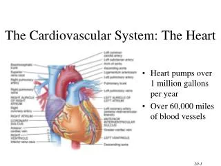
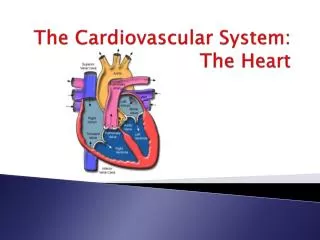
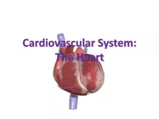
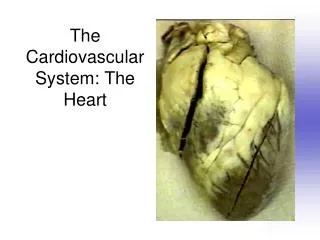
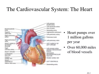
The Heart Cardiovascular System - 3. for student copy. Position of the Heart. human heart is about the size of a fist lies in the thoracic cavity w/in the mediastinum (area from sternum to vertebrae, between the lungs) tilted @ angle so its inferior surface lies against the diaphragm.

E N D
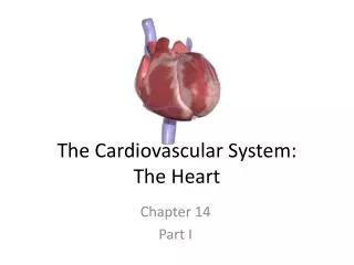
The HeartCardiovascular System - 3 for student copy
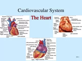
Position of the Heart • human heart is about the size of a fist • lies in the thoracic cavity w/in the mediastinum (area from sternum to vertebrae, between the lungs) • tilted @ angle so its inferior surface lies against the diaphragm
Parts of the Heart • Base of the heart is its superior border • Apex of the heart is lowest point
Major Heart Structures:the Pericardium • Outer Layer: Fibrous Pericardium • tough, attaches to diaphragm • Inner Layer: Serous Pericardium • dbl membrane: • outer parietal: attaches to fibrous pericardium • inner visceral layer: covers cardiac muscle • between the 2: pericardial cavity filled with serous fluid
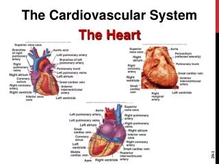
Wall of the Heart • 3 layers • outer epicardium • same as visceral pericardium • middle myocardium • cardiac muscle • inner endocardium • thin layer of endothelium that lines inside chambers of the heart & valves
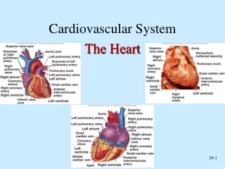
Surface Features of the Heart • 4 chambers of heart: • 2 atria form the base • Auricles (ear-like) pouch-like extensions • tip of left ventricle forms the apex • Sulci: grooves where coronary blood vessels & adipose tissue that externally mark the boundaries between the 4 heart chambers • coronary sulcus: separates atria from ventricles • anterior & posterior interventricularsulcus: separate 2 ventricles
Right Atrium • Receives deoxygenated blood from SVC & IVC
Right Atrium • inside surface has honeycombed appearance & ridges called pectinate muscles • wall separating rt & lt atrium= interatrial septum: in fetus hole called foramen ovale (blood shunts from rt atrium lt atrium avoiding pulmonary circulation); when scarred over called fossaovalis
Right Ventricle • receives blood from right atrium • sends blood to pulmonary trunk lungs to be oxygenated
Right Ventricle • inside has ridges of muscles called trabeculaecarnae: largest ones called papillary muscles: have string-like cords called cordaetendinae
Right Ventricle • separated from left ventricle by: interventricular septum
Left Atrium • receives oxygenated blood thru 4 pulmonary veins • delivers blood to left ventricle • seen on posterior surface of heart
Left Atrium • thin-walled • identifiable characteristic: 4 pulmonary veins entering it
Left Ventricle • receives oxygenated blood from left atrium • sends blood to systemic circulation thru Aorta • has thickest muscle (pumps blood the farthest)
4 Heart Valves • control 1-way flow of blood • 2 AV valves • between atria & ventricles • Tricuspid : rt AV valve • Mitral : lt AV valve, aka bicuspid • 2 semilunar valves • blood exits rt ventricle thru Pulmonary (semilunar) valve • blood exits lt ventricle thru Aortic (semilunar) valve
AV Valves • Tricuspid valve • Mitral Valve
Semilunar Valves • Pulmonary Valve • Aortic Valve
Blood Flow thru the Heart • thinner walled atria receive blood returning to heart from veins • pressure of blood in filled atria opens the AV valves & most of the blood flows into ventricles • both atria contract simultaneously to pump remaining blood into ventricles
Blood Flow thru the Heart • when atria have stopped contracting AV valves close • Ventricles contract together forcing semilunar valves open • walls of left ventricle thicker providing more force to pump blood thru systemic circulation
Blood Flow thru the Heart • Ventricular Systole: • when both ventricles are contracting • AV valves close • Semilunar valves open • Ventricular Diastole: • when both ventricles relaxed • Semilunar valves close • AV valves open
Heart Sounds • Auscultation: listening to body sounds • 1 heartbeat produces 2 heart sounds: lub-dub • heart murmurs: abnl heart sounds usually due to valve abnl • http://www.blaufuss.org/tutorial/index1.html
Pulse • when ventricles contract a blood pressure wave is produced that travels in the arteries and can be felt as your pulse • radial pulse: check over radial artery • carotid artery pulse: check over carotid artery
Calculate Pulse • Count the # of beats in 15 s and multiply x 4 • If the math is too difficult count for 30 s and multiple x 2
Blood Pressure • pressure exerted by blood against blood vessel walls • highest in the aorta & large elastic arteries & decreases as arteries get smaller & further from heart
Systolic Blood Pressure • top # on a BP • pressure generated by ventricular systole • normal adult: ~120
Diastolic BP • bottom # on BP • pressure exerted during ventricular diastole • normal adult: 60- 80
Arterial Blood Pressure • normal adult ~ 120/80 • normal venous BP: ~16 mm Hg
BP • pump used to inflate cuff to a pressure > the systolic pressure: • puts pressure on the artery, flattens it, & stops blood flow in the artery • pressure slowly released from cuff as stethoscope used to auscultate over brachial artery
BP • reported in mm Hg • as pressure in cuff becomes < pressure in artery…examiner will hear a sound can be heard, caused by the turbulent flow of blood as artery goes from flattened normal