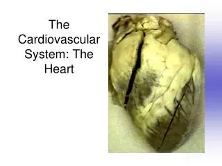
The Cardiovascular System: The Heart
480 likes | 835 Vues
The Cardiovascular System: The Heart. Heart Anatomy. Approximately the size of your fist Location Superior surface of diaphragm Left of the midline Anterior to the vertebral column, posterior to the sternum. Heart Anatomy. Figure 18.1. Coverings of the Heart: Anatomy.

The Cardiovascular System: The Heart
E N D
Presentation Transcript
Heart Anatomy • Approximately the size of your fist • Location • Superior surface of diaphragm • Left of the midline • Anterior to the vertebral column, posterior to the sternum
Heart Anatomy Figure 18.1
Coverings of the Heart: Anatomy • Pericardium – a double-walled sac around the heart that protects the heart and anchors it to the diaphragm, sternum and major blood vessels attached to the heart • Epicardium: attached to surface of the heart. Inner layer of pericardium – protects the heart • Endocardium: membrane forming the inner layer of the heart wall. Lines inner spaces of the heart • Myocardium: bulk of heart wall – cardiac muscle tissue
Pericardial Layers of the Heart Figure 18.2
Heart Wall • The clear tissue being lifted up by the scalpel is the pericardium
External Heart: Major Vessels of the Heart (Anterior View) • Vessels returning blood to the heart include: • Superior and inferior venae cavae • Right and left pulmonary veins • Vessels conveying blood away from the heart include: • Pulmonary trunk, which splits into right and left pulmonary arteries • Ascending aorta (three branches) – brachiocephalic, left common carotid, and subclavian arteries
External Heart: Anterior View Figure 18.4b
Superficial Heart Anatomy • When not filled with blood, the outer portion of each atrium deflates and becomes a lumpy, wrinkled flap. • This extension is called the auricle (looks like an external ear). • The coronary sulcus, marks the boundary between the atria and ventricles. • The interventricular sulci are shallow depressions that mark the boundary between left and right ventricles. • The attached top is the base and the apex is the inferior tip.
External Heart: Major Vessels of the Heart (Posterior View) • Vessels returning blood to the heart include: • Right and left pulmonary veins • Superior and inferior venae cavae • Vessels conveying blood away from the heart include: • Aorta • Right and left pulmonary arteries
Superior Vena Cava. Brings blood from the head, neck and shoulders to the right atrium Interatrial septum Inferior Vena Cava Brings blood back to the right atrium from the rest of the body
External Heart: Posterior View Figure 18.4d
Gross Anatomy of Heart: Frontal Section Figure 18.4e
1 2 3 Chordae tendineae Papillary muscle
Interventricular septum
Pathway of Blood Through the Heart and Lungs • Right atrium tricuspid valve right ventricle pulmonary semilunar valve pulmonary arteries (These arteries are the only arteries in the body that carries oxygen poor blood.) lung (re-oxygenation and removal of carbon dioxide.) pulmonary veins left atrium bicuspid valve left ventricle aortic semilunar valve aorta systemic circulation http://www.medtropolis.com/VBody.asp
Ventricular Differences • The anatomical differences between the right and left ventricles are as follows: The right ventricle is relatively thin. The left ventricle has a massive muscular wall. • WHY?????????
Left Ventricle Heart Wall Right Ventricle Heart Wall
Pathway of Blood Through the Heart and Lungs Rhythmic contractions of the heart wall propelling blood through the body. Figure 18.5
Coronary Circulation: Arterial Supply Figure 18.7a
Coronary Circulation: Venous Supply Figure 18.7b
Heart Valves • Heart valves ensure unidirectional blood flow through the heart • Atrioventricular (AV) valves lie between the atria and the ventricles • AV valves prevent backflow into the atria when ventricles contract • Chordae tendineae anchor AV valves to papillary muscles
Heart Valves Figure 18.8a, b
Heart Valves Figure 18.8c, d
Heart sounds • Heart Sounds: “lub-dup” – first lub is the long deep booming of blood hitting the large AV valves as they close. (ventricular contraction). Second dup is the short snapping of the closing of the smaller SL valves (ventricular relaxation) http://www.med.ucla.edu/wilkes/inex.htmhttp://depts.washington.edu/physdx/heart/demo.html http://www.blaufuss.org/
Electrical System of the heart • heart receives impulses from the autonomic nervous system. • system of specialized cells that send electrical impulses to each cardiac muscle cell. • http://www.cellsalive.com/myocyte.htm • pace setting cell: sets the rate for every cardiac muscle cell in the heart. The main site of these specialized cells is located in the wall of the R atrium called the sinoatrial node.
http://www.nhlbi.nih.gov/health/dci/Diseases/hhw/hhw_electrical.htmlhttp://www.nhlbi.nih.gov/health/dci/Diseases/hhw/hhw_electrical.html
EKG –Electrocardiogram: Recording of electrical changes that occur in the heart during a cardiac cycle.
P wave: SA node fires sending a wave of electrical activity through the atria causing depolarization. • QRS complex:: The impulse reaches the ventricles causing them to depolarize. The thick muscles of the ventricles cause 3 waves. • T wave: Repolarization of the ventricular myocardium. The ventricles are relaxing and preparing for the next contraction. (atrial repolarization is masked by the QRS waves)
Blood Vessels • Arteries and Arterioles: three walled tubes that transport blood away from the heart and maintain blood pressure. As they extend away from the heart they become smaller and branch off. The arterioles are the thinner walled vessels averaging .5mm in diameter help in regulation of peripheral resistance.
Capillaries: microscopic vessels with single layer of cells making up the walls. Exchange site (nutrients and gasses) between blood and interstitial fluid. • Venules and Veins: three walled tubes that carry blood toward the heart. Thinner than arteries. By the time blood reaches the venules, the pressure is low. • Valves: Veins of limbs have one way valves allowing the blood to flow only in one direction. • Varicose veins enlarged veins = swollen and raised on skin surface dark purple or blue, and look twisted and bulging. Commonly found on the backs of the calves or on the inside of the leg. Develop when valves in the veins stop working properly. As a result, blood pools in the veins and causes them to get larger.
http://www.heartsite.com/html/the_heart_3.html (student tutorial they can go to for additional help) Angina pectoris: oxygen supply reduced to a point of damaging cardiac muscle cells but not killing them. Symptoms – chest pain, tightness in chest, labored breathing, dizziness, weakness. Myocardial infarction: Cardiac muscle cell death due to an interruption in blood flow. Cardiac arrest: complete stoppage of the heart
http://www.heartpoint.com/arrhythmias-ventricular.html • http://www.heartpoint.com/afibgallery.html • http://www.heartpoint.com/coronartdisease.html • http://www.heartpoint.com/angioplasty.html • http://www.heartpoint.com/coumadin.html • http://heartpoint.com/pacerintro.html
BELOW WEB SITES FOR VIRTUAL HEART SURGERY http://www.pbs.org/wgbh/nova/eheart/transplant.html (simplified) http://www.abc.net.au/science/lcs/heart.htm (best site) Tutorials with quizes: http://highered.mcgraw-hill.com/sites/0072495855/student_view0/chapter22/animation__the_cardiac_cycle__quiz_1_.html http://highered.mcgraw-hill.com/sites/0072495855/student_view0/chapter22/animation__the_cardiac_cycle__quiz_2_.html beating heart surgery Surgeon describes what he is doing. http://www.youtube.com/watch?v=Zxqj1BcBpIg silent surgery (Great short video of actual surgery) http://www.youtube.com/watch?v=HW-6SlIako0&feature=related (surgeon describes as heart stops for procedure, and is restarted after surgery is completed.) Oen Heart Surgery.webloc
BLOOD PRESSURE: • force exerted by blood against the walls of vessels. • First number is systolic pressure – pressure when heart is contracting. A large volume of blood pumps into the aorta. The walls of the arteries stretch to accommodate the large volume in a small space: maximum pressure.
Second number is the diastolic pressure. Resting pressure. It occurs when the aorta has emptied and the pressure is at a minimum. • Sphygmomanometer: an instrument used to measure arterial blood pressure. The cuff is wrapped around the arm over the brachial artery. • Pulse: palpation of heart rate.