Download
1 / 112
1.13k likes | 1.28k Vues
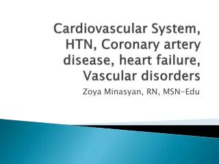
Cardiovascular System, HTN, Coronary artery disease, heart failure, Vascular disorders. Zoya Minasyan , RN, MSN- Edu. Blood Flow Through the Heart. Arrows indicate direction of flow .

E N D
Cardiovascular System, HTN, Coronary artery disease, heart failure, Vascular disorders ZoyaMinasyan, RN, MSN-Edu
Blood Flow Through the Heart Arrowsindicate direction of flow. 1, The right atrium receives venous blood from the inferior and superior vena cava and the coronary sinus. The blood then passes through the tricuspid valve into the right ventricle. 2, With each contraction, the right ventricle pumps blood through the pulmonic valve into the pulmonary artery and to the lungs. 3, Oxygenated blood flows from the lungs to the left atrium by way of the pulmonary veins. 4, It then passes through the mitral valve and into the left ventricle. 5, As the heart contracts, blood is ejected through the aortic valve into the aorta and thus enters the systemic circulation.
Cardiac Valves Anatomic structures of the heart and heart valves.
Coronary Arteries and Veins Coronary arteries and veins.
Common Sites for Palpating Arteries Common sites for palpating arteries. 6
Mechanical System • Systole: Contraction of myocardium • Diastole: Relaxation of myocardium • Cardiac output: Amount of blood pumped by each ventricle in 1 minute CO = SV × HR • Preload • Volume of blood in ventricles at the end of diastole • Afterload • Peripheral resistance against which the left ventricle must pump
Relationship of Electrocardiogram, Cardiac Cycle, and Heart Sounds 8
Chest X-ray Standard posterior-anterior view.
Diagnostic Studies of Cardiovascular System • Noninvasive studies • Magnetic resonance imaging • Computed tomography • Echocardiogram • Nuclear cardiology
Diagnostic Studies of Cardiovascular System • MRI. magnetic resonance imaging can detect and localize areas of MI in a three-dimensional view and can assist in the final diagnosis of MI. It is also plays a role in prediction of recovery from MI and in the diagnosis of congenital heart and aortic disorders. • Computed Tomography Angiography (CTA) • noninvasive scan used to quantify calcium deposits in coronary arteries. • Electron beam computed tomography (EBCT), also known as ultrafast CT, uses a scanning electron beam to quantify calcification in the coronary arteries and heart valves. Currently, EBCT testing is used primarily for risk assessment in asymptomatic patients and to assess for heart disease in patients with atypical symptoms potentially due to cardiac causes. • Echocardiogram. The echocardiogram uses ultrasound waves to record the movement of the structures of the heart and provides information about abnormalities of • valvularstructure and motion, • cardiac chamber size and contents, • ventricular muscle and septal motion and thickness, • pericardial sac, and • ascending aorta. • Nuclear Cardiology. One of the most common nuclear imaging tests is the multigated acquisition (MUGA) or cardiac blood pool scan. This test provides information on wall motion during systole and diastole, cardiac valves, and EF.
Echocardiogram Apical four-chamber two-dimensional echocardiographic view in a normal patient. LA, Left atrium; LV, left ventricle; MV, mitral valve; RA, right atrium; RV, right ventricle; TV, tricuspid valve.
Computed Tomography Examples of coronary calcification of the left anterior descending coronary artery (large arrow) and left circumflex artery (small arrow) as seen on electron beam computed tomography.
Diagnostic Studies of Cardiovascular System • Invasive studies • Cardiac catheterization and coronary angiography • Intracoronary ultrasound • Fractional flow reserve • Electrophysiology study • Blood flow and pressure measurements
Diagnostic Studies of Cardiovascular System • Cardiac Catheterization. It provides information about CAD, coronary spasm, congenital and valvular heart disease, and ventricular function. Cardiac catheterization is also used to measure intracardiac pressures and O2 levels, as well as CO and EF. • Intracoronary Ultrasound.(ICUS), also known as intravascular ultrasound (IVUS), is an invasive procedure performed in the catheterization laboratory in conjunction with coronary angiography. The 2-D or 3-D ultrasound images provide a cross-sectional view of the arterial walls of the coronary arteries. • Fractional Flow Reserve.(FFR) is a procedure that is done during a cardiac catheterization. It involves using a special wire that can measure pressure and flow in the coronary artery. • Electrophysiologic Study.(EPS) is the direct study and manipulation of the electrical activity of the heart using electrodes placed inside the cardiac chambers. It provides information on SA node function, AV node conduction, and ventricular conduction. • Blood Flow and Pressure Measurements. • Peripheral Vessel Blood Flow. Duplex imaging is useful in the diagnosis of occlusive disease in the peripheral blood vessels and for the diagnosis of thrombophlebitis. Peripheral vessel blood flow is assessed by injecting contrast media into the appropriate arteries or veins (arteriography or venography). • Hemodynamic Monitoring. Bedside hemodynamic monitoring of pressures of the cardiovascular system is frequently used to assess cardiovascular status and monitor patient response to interventions. Invasive hemodynamic monitoring using intraarterial and pulmonary artery catheters can be used to monitor arterial BP, intracardiac pressures, and CO.
Normal Left Coronary Artery Angiogram Normal left coronary artery angiogram. 16
Question A patient arrives at an urgent care center after experiencing unrelenting substernal and epigastric pain and pressure for about 12 hours. The nurse reviews laboratory results with the understanding that at this point in time, a myocardial infarction would by indicated by peak levels of: 1. Troponin T. 2. Homocysteine. 3. Creatine kinase-MB. 4. Type b natriuretic peptide.
Cardiac markers • Troponin is the biomarker of choice in the diagnosis of myocardial infarction. Troponin is a myocardial muscle protein released into the circulation within 1 hr after injury. Troponin levels peak at 10 to 24 hours. • When Myocardial cells are injured, they release their content, including enzymes and other proteins, into the circulation • Enzymes called creatinekinase (CK); lactate dehdrogenaze-(LDH); and serum aspartateaminotransferase (AST) formally called glutamic-oxaloacetictransaminase (SGOT) • CK is present in heart ,brain and skeletal muscles • CK-MM primarily in skeletal muscle • CK-BB brain and nervous tissue • Ck-MB specific for myocardial injury; • It rises in 4-6 after onset, peak in 18-24 hr
Gerontologic Consideration • Age alters the cardiovascular response to physical and emotional stress. • Heart valves become thick and stiff. • Frequent need for pacemakers • Less sensitive to β-adrenergic agonist drugs • Increase in SBP; decrease or no change in DBP
HypertensionDefinition • Persistent elevation of • Systolic blood pressure ≥140 mm Hg OR Diastolic blood pressure ≥90 mm Hg OR • Current use of antihypertensive medication(s) • Table 33-6 page 769 20
BP classification • Normal <120and <80 • Prehypertension 120-139 and 80-89 • Stage one hypertention 140-159 and 90-99 • Stage 2 hypertention >or=160 and >or=100
Factors Influencing Blood Pressure (BP) Systemic Vascular Resistance Blood Pressure Cardiac Output = × 22
Etiology of Hypertension • Primary (essential) hypertension • Contributing factors • ↑ SNS activity • ↑ Sodium-retaining hormones and vasoconstrictors • Diabetes mellitus • > Ideal body weight • ↑ Sodium intake • Excessive alcohol intake
Etiology of Hypertension • Secondary hypertension • Contributing factors • Aortic abnormalities • Renal disease • Endocrine disorders • Neurologic disorders • Cirrhosis • Sleep apnea
Risk Factors for Primary Hypertension • Age • Alcohol • Cigarette smoking • Diabetes mellitus • Elevated serum lipids • Excess dietary sodium • Gender • Family history • Obesity • Ethnicity • Sedentary lifestyle • Socioeconomic status • Stress
Factors Influencing BP Hypertension develops when one or more of the BP regulating mechanisms are defective. EDRF, Endothelium-derived relaxing factor.
Pathophysiology of Primary Hypertension • Insulin resistance and hyperinsulinemia • High insulin concentration stimulates SNS activity and impairs nitric oxide–mediated vasodilation • Stress and increased SNS activity • Produce increased vasoconstriction • ↑ HR • ↑ Reninrelease(first-vasoconstrictor, second -can cause the stimulation of aldosterone which causes Na and H2O retention) • Altered renin-angiotensin mechanism: High plasma reninactivity • Endothelial cell dysfunction • Water and sodium retention • High sodium intake may activate a number of pressor mechanisms, resulting in water retention.
HypertensionComplications • Target organ diseases occur most frequently in the • Heart • Brain- Cerebrovasculardisease-Stroke • Peripheral vasculature • Kidney: Nephrosclerosis • Eyes Retinal damage
Hypertension: Collaborative Care • Lifestyle modifications • Weight reduction: Weight loss of 10 kg (22 lb) may decrease SBP by approx 5 to 20 mm Hg • Dietary sodium reduction: <2300 mg of sodium/day • Moderation of alcohol consumption: • Men: No more than 2 drinks/day • Women: No more than 1 drink/day • Physical activity: Regular physical (aerobic) activity, at least 30 minutes, most days of the week • Avoidance of tobacco products • Psychosocial risk factors • Drug therapy: Classifications of drugs used to treat hypertension • Diuretics • Adrenergic inhibitors • Direct vasodilators • Angiotensin-converting enzyme inhibitors • Angiotensin II receptor blockers • Calcium channel blockers
Question A patient’s blood pressure has not responded consistently to prescribed medications for hypertension. The first cause of this lack of responsiveness the nurse should explore is: 1. Progressive target organ damage. 2. The possibility of drug interactions. 3. The patient not adhering to therapy. 4. The patient’s possible use of recreational drugs.
Hypertensive Crisis • Severe increase in BP (>220/140) • Often occurs in patients with a history of HTN who have failed to comply with medications or who have been undermedicated • Hypertensive emergency = Evidence of acute target organ damage: • Hypertensive encephalopathy, cerebral hemorrhage • Acute renal failure • Myocardial infarction • Heart failure with pulmonary edema
Hypertensive CrisisNursing and Collaborative Management • Hospitalization • IV drug therapy • Monitor cardiac and renal function • Neurologic checks • Determine cause • Education to avoid future crises
Coronary Artery Disease and Acute Coronary Syndrome • Atherosclerosis: Type of blood vessel disorder • Begins as soft deposits of fat that harden with age • Referred to as “hardening of arteries” • Greek words: athere, meaning “fatty mush,” and skleros, meaning “hard.” • Terms to describe the disease process • Arteriosclerotic heart disease • Cardiovascular heart disease • Coronary artery disease (CAD)
Coronary Artery Disease Etiology and Pathophysiology • Atherosclerosis is the major cause of CAD. • Characterized by a focal deposit of cholesterol and lipid, primarily within the intimal wall of the artery • Endothelial lining altered as a result of inflammation and injury • C-reactive protein (CRP) • Nonspecific marker of inflammation • Increased in many patients with CAD • Chronic exposure to CRP associated with unstable plaques and oxidation of LDL cholesterol
Pathogenesis of Atherosclerosis A, Damaged endothelium. B, Diagram of fatty streak and lipid core formation. C, Diagram of fibrous plaque. Raised plaques are visible: some are yellow, others are white. D, Diagram of complicated lesion: thrombus is red, collagen is blue. Plaque is complicated by red thrombus deposition.
Coronary Artery Disease :Etiology and Pathophysiology • Developmental stages: • Fatty streaks • Earliest lesions • Characterized by lipid-filled smooth muscle cells • Potentially reversible • Fibrous plaque • Beginning of progressive changes in the arterial wall • Lipoproteins transport cholesterol and other lipids into the arterial intima. • Fatty streak is covered by collagen, forming a fibrous plaque that appears grayish or whitish. • Result = Narrowing of vessel lumen • Complicated lesion • Continued inflammation can result in plaque instability, ulceration, and rupture. • Platelets accumulate and thrombus forms. • Increased narrowing or total occlusion of lumen
Risk Factors for CAD • Modifiable risk factors • Elevated serum lipids • Hypertension • Tobacco use • Physical inactivity • Non modifiable risk factors • Age • Gender • Ethnicity • Family history • Genetic predisposition
Risk factors • Serum cholesterol level greater than 200 mg/dL(5.2 mmol/L) or a fasting triglyceride level greater than 150 mg/dL (3.7 mmol/L). • Hypertension, which is defined as a BP > 140/90 mm Hg or >130/80 mm Hg if the patient has diabetes or chronic kidney disease. • Tobacco use. tobacco smoking decreases estrogen levels, placing premenopausal women at greater risk for CAD. Risk is proportionate to the number of cigarettes smoked. • Physical inactivity • Obesity is defined as a body mass index (BMI) of >30 kg/m2 and a waist circumference ≥40 inches for men and ≥35 inches for women. • 2 to 4 times greater among persons who have diabetes. • Specific psychological risk factors thought to increase risk of CAD. These include depression, acute and chronic stress, anxiety, hostility and anger, and lack of social support.
Risk Factors for CADHealth Promotion • Health-promoting behaviors • Physical fitness • 30 minutes >5 days/week • Regular physical activity contributes to • Weight reduction • Reduction of >10% in systolic BP • In some men more than women, increase in HDL cholesterol
Risk Factors for CAD: Health Promotion • Therapeutic lifestyle changes include • a decrease in saturated fat and cholesterol and an increase in complex carbohydrates (e.g., whole grains, fruit, vegetables). • Fat intake should be about 30% of calories, with most coming from monounsaturated fats found in nuts and oils such as olive or canola oil. • For individuals without CAD, the AHA recommends eating • fatty fish twice a week because fatty fish such as salmon and tuna contain two types of omega-3 fatty acids: Eicosapentaenoic acid (EPA) and docosahexaenoic acid (DHA). Patients with CAD are encouraged to take EPA and DHA supplements with their diet. • tofu, other forms of soybean, canola, walnut, and flaxseed, because these products contain alpha-linolenic acid, which becomes omega-3 fatty acid in the body.
Risk Factors for CAD: Health Promotion • Health-promoting behaviors • Antiplatelet therapy • ASA • Clopidogrel (Plavix)
Clinical Manifestations of CAD Chronic Stable Angina • Etiology and pathophysiology • Reversible (temporary) myocardial ischemia = Angina (chest pain) • O2 demand > O2 supply • Primary reason for insufficient blood flow is narrowing of coronary arteries by atherosclerosis. • Referred pain in left shoulder and arm is from transmission of the pain message to the cardiac nerve roots. • Intermittent chest pain that occurs over a long period with the same pattern of onset, duration, and intensity of symptoms • Pain usually lasts 3 to 5 minutes. • Subsides when the precipitating factor is relieved • Pain at rest is unusual. • ECG reveals ST-segment depression and/or T-wave inversion.
Chronic Stable Angina Types of Angina • Silent ischemia • Ischemia that occurs in the absence of any subjective symptoms • Associated with diabetic neuropathy • Confirmed by ECG changes • Nocturnal angina • Occurs only at night but not necessarily during sleep • Angina decubitus • Chest pain that occurs only while lying down • Relieved by standing or sitting
Chronic Stable AnginaTypes of Angina • Prinzmetal’s (variant) angina • Occurs at rest usually in response to spasm of major coronary artery • Seen in patients with a history of migraine headaches and Raynaud’s phenomenon • Spasm may occur in the absence of CAD. • May occur during REM sleep • May be relieved by moderate exercise or may disappear spontaneously • Microvascular angina • May occur in the absence of significant coronary atherosclerosis or coronary spasm • Pain is related to myocardial ischemia associated with abnormalities of the coronary microcirculation. • Coronary microvascular disease
Chronic Stable Angina Nursing and Collaborative Management • Drug therapy: Goal: ↓ O2 demand and/or ↑ O2 supply • Short-acting nitrates: • Sublingual • Long-acting nitrates • Nitroglycerin (NTG) ointment • Transdermal controlled-release NTGβ-Adrenergic blockers • Calcium channel blockers • If β-adrenergic blockers are poorly tolerated, contraindicated, or do not control angina • Used to manage Prinzmetal’s angina • Angiotensin-converting enzyme inhibitors
Chronic Stable AnginaNursing and Collaborative Management • Diagnostic studies • Health history/physical examination • Laboratory studies • 12-lead ECG • Chest x-ray • Echocardiogram • Exercise stress test • Cardiac catheterization/coronary angiography • Coronary revascularization: Percutaneous coronary intervention (PCI) • Balloon angioplasty • Stent
Placement of a Coronary Artery Stent Fig. 34-6. Placement of a coronary artery stent. A, The stent is positioned at the site of the lesion. B, The balloon is inflated, expanding the stent. The balloon is then deflated and removed. C, The implanted stent is left in place.
Pre- and Post- Percutaneous coronary intervention (PCI) With Stent Placement Fig. 34-7. A, A thrombotic occlusion of the right coronary artery is noted (arrows).B, Right coronary artery is opened and blood flow restored following angioplasty and placement of a 4-mm stent.
Acute Coronary Syndrome • When ischemia is prolonged and is not immediately reversible, acute coronary syndrome (ACS) develops. • Clinical Manifestations of ACS • Unstable Angina • Change in usual pattern • New in onset • Occurs at rest • Has a worsening pattern • UA is unpredictable and represents a medical emergency. • Result of sustained ischemia (>20 minutes), causing irreversible myocardial cell death (necrosis) • Necrosis of entire thickness of myocardium takes 4 to 6 hours.
Acute Myocardial Infarction Fig. 34-10. Acute myocardial infarction in the postero-lateral wall of the left ventricle. This is demonstrated by the absence of staining in the areas of necrosis (white arrow).Note the scarring from a previous anterior wall myocardial infarction (black arrow).