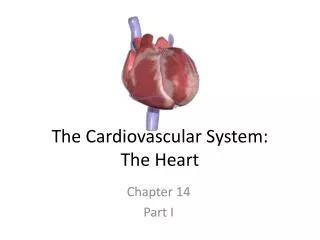
The Cardiovascular System: The Heart
380 likes | 1.18k Vues
The Cardiovascular System: The Heart. Chapter 14 Part I. Objectives for Chaper 14. To understand the: Structure and function of the heart Physiology underlying the cardiac cycle Generation of electrical impulses Use and interpretation of the electrocardiogram. Cardiac Output (CO).

The Cardiovascular System: The Heart
E N D
Presentation Transcript
The Cardiovascular System:The Heart Chapter 14 Part I
Objectives for Chaper 14 To understand the: • Structure and function of the heart • Physiology underlying the cardiac cycle • Generation of electrical impulses • Use and interpretation of the electrocardiogram
Cardiac Output (CO) • Volume of blood pumped/min by each ventricle • Stroke volume (SV) = blood pumped/beat by each ventricle • Heart rate(HR)= the number of beats/minute • CO = SV x HR • Total blood volume = about 5.5L
Regulation of Cardiac Rate • Without neuronal influences – SA node will drive heart at rate of its spontaneous activity • Chronotropic effect– normally sympathetic and parasympathetic activity influence HR • Main controller of HR – autonomic innervation of SA node • Sympathetic and Parasympathetic nerve fibers modify rate of spontaneous depolarization
Regulation of Cardiac Rate • Effect of autonomic nerves on the pacemaker potentials in the SA node • NE and Epi stimulate opening of pacemaker HCN channels • This depolarizes SA node faster, increasing HR • ACh promotes opening of K+ channels • The resultant K+ outflow counters Na+ influx, slowing depolarization and decreasing HR
Cardiac Control Center • In medulla oblongata coordinates activity of autonomic innervation • Sympathetic endings in atria and ventricles can stimulate increased strength of contraction
Stroke Volume • Determined by 3 variables: • End diastolic volume(EDV) = volume of blood in ventricles at end of diastole • Total peripheral resistance(TPR) = resistance to blood flow in arteries • Contractility = strength of ventricular contraction
Regulation of Stroke Volume • EDV – workload (preload) on heart prior to contraction • SV is directly proportional to preload and contractility • Strength of contraction varies directly with EDV • Total peripheral resistance = afterload which impedes ejection from ventricle • Ejection fraction = SV/ EDV • Normally is about 60% • Useful clinical diagnostic tool
Frank-Starling Law of the Heart • As EDV is increased, the stroke volume is increased • States that strength of ventricular contraction varies directly with EDV • Intrinsicpropertyof myocardium • As EDV increases, myocardium is stretched more • causing greater contraction and SV
Frank-Starling Law of the Heart (a) State of myocardial sarcomeres just before filling • Actins overlap, actin-myosin interactions are reduced and contraction would be weak (b, c, d) Increasing interaction of actin and myosin • allowing more force to be developed
Extrinsic Control of Contractility • At any given EDV – contraction depends upon level of sympathoadrenal activity • NE and Epi produce increase in HR and contraction (positive inotropic effect) • Due to increased Ca2+ in sarcomeres
The Regulation of Cardiac Output • Solid lines – Factors that stimulate CO • Dashed arrows – Factors that inhibit CO
Venous Return • Return of blood to the heart via veins • Controls EDV and thus SV and CO • Dependent on: • Blood volume and venous pressure • Sympathetic vasoconstriction • Skeletal muscle pumps • Pressure drop during inhalation
Venous Return • Veins hold most of the body’s blood (~70%) • called capacitance vessels • Have thin walls, stretch easily to accommodate more blood • without increased pressure (higher compliance) • only 0-10 mm Hg pressure
Blood Volume • Constitutes small fraction of total body fluid • 2/3 of body H2O – inside cells (intracellular compartment) • 1/3 total body H2O – in extracellular compartment • 80% of this is interstitial fluid; 20% is blood plasma
Exchange of Fluid between Capillaries and Tissues • Distribution of ECF between blood and interstitial compartments is in state of dynamic equilibrium • Movement out of capillaries is driven by hydrostatic pressure exerted against capillary wall • Promotes formation of tissue fluid • Net filtration pressure= hydrostatic pressure in capillary (17-37 mm Hg) - hydrostatic pressure of ECF (1 mm Hg)
Exchange of Fluid between Capillaries and Tissues • Movement also affected by colloid osmotic pressure • = osmotic pressure exerted by proteins in fluid • Difference between osmotic pressures in and outside of capillaries (oncotic pressure) affects fluid movement • Plasma osmotic pressure = 25 mm Hg; interstitial osmotic pressure = 0 mm Hg
Overall Fluid Movement • Determined by net filtration pressure and forces opposing it (Starling forces) • (Pc + I)– (Pi + p) • [fluid out] – [fluid in] • Pc = Hydrostatic pressure in capillary • i = Colloid osmotic pressure of interstitial fluid • Pi = Hydrostatic pressure in interstitial fluid • p = Colloid osmotic pressure of blood plasma
Distribution of Fluid Across Capillary Walls • Tissue, or interstitial fluid, formed by filtration (yellow arrows) • result of BPs at the arteriolar ends of capillaries • Returned to venular ends by the colloid osmotic pressure of plasma proteins
Edema • Normally filtration, osmotic reuptake, and lymphatic drainage maintain proper ECF levels • Edema – excessive accumulation of fluid resulting from: • High arterial blood pressure, increases capillary pressure causes excessive filtration • Venous obstruction produces a congestive increase in capillary pressure • Leakage of plasma proteins into interstitial fluid causes reduced osmotic flow of water into capillaries • Myxedema (excess production of glycoproteins in extracellular matrix) from hypothyroidism • Low plasma protein levels resulting from liver disease • Obstruction of lymphatic drainage
Elephantiasis • Parasitic larvae – block lymphatic drainage • Produces tissue edema • with tremendous enlargement of limbs and scrotum
Regulation of Blood Volume by Kidney • Urine formation begins with filtration of plasma in glomerulus • Filtrate passes through and is modified by nephron • Volume of urine excreted can be varied by changes in reabsorption of filtrate • Adjusted according to needs of body by action of hormones
ADH (Vasopressin) • Released by posterior pituitary – osmoreceptors in hypothalamus detect high osmolality • From excess salt intake or dehydration • Causes thirst • Stimulates H2O reabsorption from urine • Homeostasis maintained by these countermeasures
Aldosterone • Steroid hormone secreted by adrenal cortex • Helps maintain blood volume and pressure through reabsorption and retention of salt and water • Release stimulated by salt deprivation, low blood volume, and pressure
Renin-Angiotension-Aldosterone System • When there is a salt deficit, low blood volume, or pressure, angiotensin IIis produced • causes a number of effects all aimed at increasing blood pressure: • Vasoconstriction, aldosterone secretion, thirst
Angiotensin II • Renin-angiotensin-aldosterone system helps to maintain homeostasis • Through negative feedback control of blood volume and pressure • JGA secretes renin cleaves angiotensinogen to angiotensin I (10 aa) • ACE (angiotensin-converting enzyme) removes 2 aa angiotensin II
Atrial Natriuretic Peptide (ANP) • Expanded blood volume is detected by stretch receptors in left atrium • causes release of ANP • ANP inhibits aldosterone, promoting salt and water excretion to lower blood volume • And promotes vasodilation
ANP • Together with decreased ADH (neg feedback) lowers blood volume and venous return