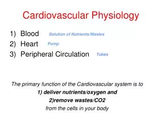
Cardiovascular Physiology
Cardiovascular Physiology. Cardiovascular disease is #1 cause of death Major underlying cause is ischemia due to: atherosclerosis (plaquing) white thrombus red thrombus artery spasm.


Cardiovascular Physiology
E N D
Presentation Transcript
Cardiovascular Physiology • Cardiovascular disease is #1 cause of death • Major underlying cause is ischemia due to: atherosclerosis (plaquing) white thrombus red thrombus artery spasm
It ain't what you don't know that gets you into trouble. It's what you know for sure that just ain't so. Mark Twain
Connective Tissue • Ordinary • Loose connective tissue (areolar tissue) • Dense ordinary connective tissue • Regular vs. Irregular • Special • Adipose tissue (fat) • Blood cells • Blood cell forming tissue • Myeloid or lymphatic tissue • Cartilage • Bone
Events in Hemostasis • Hemostasis-prevention of blood loss • Mechanisms • vascular spasm • formation of a platelet plug • blood coagulation • fibrous tissue growth to seal
Hemostasis • Vascular Constriction-associated w/ trauma • neural reflexes • SNS induced constriction from pain • local myogenic spasm • responsible for most of the constriction • local humoral factors • thromboxane A2 from platelets • Spasm trauma
Platelet Plug Platelets function as whole cells but cannot divide Platelets contain: actin & myosin enzymes & calcium ADP & ATP Thromboxane A2 serotonin growth factor
Platelet Cell Membrane Contains: Glycoproteins that avoid the normal endothelium but adhere to damaged area Phospholipids containing platelet factor 3 a.k.a. thromboplastin-initiates clotting
Mechanism of Platelet Activation When platelets contact damaged area they 1) swell 2) irregular form w/ irradiating processes protruding from surface 3) contractile proteins contract causing granule release 4) secrete ADP, Thromboxane A2 & serotonin
Thromboxane A2 1) Vasoconstrictor 2) Potentiates the release of granule contents (not essential for releaseto occur)
Platelets • Important in minute ruptures • lack of platelets associated with small hemorrhagic areas under skin and throughout internal tissues • half-life of 8-12 days • eliminated primarily by macrophage action • (greater than 1/2 of all macrophages in spleen) • 150,000-300,000 per l
Role of Endothelium • Prevents platelet aggregation • produces PGI2 (prostacyclin)- • vasodilator • stimulates platelet adenyl cyclase which suppresses release of granules • limits platelet extension • produces factor VIII (clotting)
Prostaglandin synthesis • Phospholipid Arachidonic acid requires Lipase • Arachidonic acidPGG2-PGH2 requires fatty acid cyclooxygenase • PGG2-PGH2Thromb. A2 requires Thromb. synthetase • PGG2-PGH2PGI2 requires Prostacyclin synthetase • Aspirin and Ibuprofen block Fatty acid cyclooxygenase
Anticoagulants vs Lysis of clots • Anticoagulants • prevents clots from forming • chelators-tye up calcium (citrate, oxylate) • heparin- complexes with Antithrobin III • dicumarol-inhibition of Vit. K dependent factors • factors II, VII, IX, X (synthesized by hepatocytes • Aka cumadin, warfarin • Lysis of Clots • Plasmin (from plasminogen)
Activators of Plasminogen • Endogenous Activators • tissues • plasma • urine • Exogenous Activators • streptokinase • tPA (tissue plasminogen activator)
Aspirin & Ibuprofen • Block both thromboxane A2 & prostacyclin production by blocking fatty acid cyclooxygenase which converts arachidonic acid to PGG2 & PGH2 (intermediates) • Why take aspirin to prevent heart attacks?
Reperfusion injury • Most of the frank tissue damage associated with infarction occurs upon reperfusion • associated with the formation of highly reactive oxygen species with unpaired electrons. “free radicals” • When pressure on tissues relieved & again perfused with blood, free radicals are generated
Collateralization • The ability to open up alternate routes of blood flow to compensate for a blocked vessel • Angiogenesis • Vasodilatation • Role of the SNS ?? • May impede • May augment
Blood Coagulation- Thrombosis • Extrinsic mechanism-initiated by chemical factors released by damaged tissues • Intrinsic mechanism-requires only components in blood & trauma to blood or exposure to collagen (or foreign surface)
Clotting factors • I- fibrinogen • II- Prothrombin • III- Thromboplastin • IV- Calcium • V- Proaccelerin • VII- Serum prothombin conversion acclerator • VIII- antihemophilic factor (A)
Clotting factors (cont.) • IX- antihemophilic factor B “christmas factor” • X- Stuart factor • XI- antihemophilic factor C • XII- Hageman factor • XIII- Fibrin-stabilizing factor • Prekallikrein- Fletcher factor • High molecular weight kininogen • Platelets
Hepatocytes role in clotting • Liver synthesizes 5 clotting factors • I (fibrinogen) • II (prothrombin) • VII (SPCA) • IX (AHF B) • X (Stuart factor) • Coumarin (warfarin or cumadin) depresses liver formation of II, VII, IX, X by blocking action of vitamin K
Hemophilia • Sex linked on X chromosome • occurs almost exclusively in males • 85% of cases- defect in factor VIII • 15% of cases- defect in factor IX • varying degree of severity from mild severe
Blood Coagulation • The key step is the conversion of fibrinogen to fibrin which requires thrombin • thrombin • fibrinogen--------------->fibrin
Intrinsic pathway • Factor XII activated when blood contacts a negatively charged surface (collagen, glass) • Activated XII + Kalikrein+ Kinnogen will activate Factor XI • Activated XI + Ca++ will activate both Factors IX & VIII • Activated IX + VIII + Phospholipid + Ca++ will activate Factors X & V • Activated X + V + Phospholipid + Ca++ will convert Prothrombin to Thrombin
Extrinsic Pathway • Tissue thromboplastin + Factor VII + Ca++ will activate Factors X & V • Activated X & V + Phospholipid + Ca++ will convert Prothrombin to Thrombin
Final Common Steps • Once Fibrinogen has been converted to Fibrin by Thrombin it is changed from the soluble monomer to the insoluble polymer by the activated Factor XIII • Factor XIII is activated by Thrombin and Ca++
Lysis of Clots • Clots may be liquefied by (fibrinolysis) by a proteolytic enzyme “plasmin” • It circulates in the blood in an inactive form known as plasminogen • Activators are found in tissues, plasma, and urine • It can also be activated by exogenous activators such as tPA, or streptokinase
Risk factors in Heart Disease • Increasing age • Male gender • Heredity (including race) • Tobacco Smoke • High blood cholesterol • High blood pressure • Physical inactivity • Obesity/overweight • Diabetes Mellitus • High blood homocysteine
Homocysteine • Amino acid in the blood that may irritate blood vessels promoting atherosclerosis • Can also cause cholesterol to change into oxidized LDL • Can make blood more likely to clot • High levels in blood (> 12 mol/L) can be reduced by increasing intake of folic acid, B6 and B12
Heart muscle • Atrial & Ventricular • striated enlongated grouped in irregular anatamosing columns • 1-2 centrally located nuclei • Specialized excitatory & conductive muscle fibers (SA node, AV node, Purkinje fibers) • contract weakly • few fibrils
Syncytial nature of cardiac muscle • Syncytium = many acting as one • Due to presence of intercalated discs • low resistance pathways connecting cardiac cells end to end • presence of gap junctions
Action potentials in cardiac muscle • Duration of action potential is from .2-.3 sec • Channels • fast Na+ channels • slow Ca++/Na+ channels • K+ channels • Permeability changes • Na+ sharp increase at onset of depolarization • Ca++ increased during the plateau • K+ increased during the resting polarized state
Membrane physiology • In excitable tissue an action potential is a pulse like change in membrane permeability • In cardiac muscle permeability changes for: • Na+ • at onset of depolarization, during repolarization • Ca++ • at onset of depolarization, during repolarization • K+ • at onset of depolarization, during repolarization
Slow vs Fast cardiac cell • Relates to the channels that open during depolarization • Typical cardiac muscle have both fast Na+ channels and slow Ca++/Na+ channels that open during depolarization • Specialized excitatory cells like the SA node only slow Ca++/Na+ channels are operational during depolarization increasing depolarization time • Tetradotoxin blocks fast Na+ channels selectively changing a fast response into a slow response
Passive ion movement across cell • Considerations • Concentration gradient • high to low • Electrical gradient • opposite charge attract, like charge repel • Membrane permeability • dependant on ion channels (open or closed) • If ion channels are open, an ion will seek its Nerst equilibrium potential • concentration gradient favoring ion movement in one direction is offset by electrical gradient
Resting membrane potential (Er) • During the Er in cardiac muscle, fast Na+ and slow Ca++/Na+ are closed, K+ channels are open. • Therefore K+ ions are free to move, and when they reach their Nerst equilibrium potential, a stable Er is maintained
Na+/K+ ATPase (pump) • The Na+/K+ pump which is energy dependent operates to pump Na+ out & K+ into the cardiac cell at a ratio of 3:2 • therefore as pumping occurs, there is net loss of one + charge from the interior each cycle, helping the interior of the cell remain negative • the protein pump utilizes energy from ATP • Digitalis binds to & inhibits this pump
Ca++ exchange protein • In the cardiac cell membrane is a protein that exchanges Ca++ from the interior in return for Na+ that is allowed to enter the cell. • The function of this exchange protein is tied to the Na+/K+ pump • if the Na+/K+ pump is inhibited, function of this exchange protein is reduced & more Ca++ is allowed to accumulate in the cardiac cell contractile strength.
Refractory Period • Absolute • unable to re-stimulate cardiac cell • occurs during the plateau • Relative • requires a supra-normal stimulus • occurs during repolarization • In a Slow response cardiac muscle cell the relative refractory period is prolonged and the refractory period is about 25% longer • in AV node & bundle this serves to protect the ventricles from supra-ventricular arrhythmias
SA node • Normal pacemaker of the heart • Self excitatory nature • less negative Er • leaky membrane to Na+/CA++ • only slow Ca++/Na+ channels operational • spontaneously depolarizes at fastest rate • overdrive suppression • contracts feebly
Overdrive Suppression • If you drive a self-excitatory cell at a rate faster than its own inherent rate, you will suppress the cell’s own automaticity • Mechanism may be due to increased activity of Na+/K+ pump creating more negative Er • Cells of the AV node and purkinje system are under overdrive suppression by the SA node
AV node • Delays the wave of depolarization from entering the ventricle • allows the atria to contract slightly ahead of the ventricles (.1 sec delay) • Slow conduction velocity due to smaller diameter fibers • In absence of SA node, AV node may act as pacemaker but at a slower rate
Effect of HR on systole/diastole • As heart rate (HR) cycle length (CL) • At a resting heart rate systole (S) < diastole (D) • Both the duration of systole and diastole shorten, but diastole shortens to a greater extent • At high HR the ventricle may not fill adequately • HR of 75 BPM; CL = .8 sec. S = .3 D = .5 • HR of 150 BPM; CL = .4 sec. S = .2 D = .2 • During systole perfusion of the myocardium is restricted by the contracting cardiac muscle compressing blood vessels (especially in LV)
Cardiac Cycle • Systole • isovolumic contraction • ejection • Diastole • isovolumic relaxation • rapid inflow- 70-75% • diastasis • atrial systole- 25-30%
Onset of Ventricular Contraction • Isovolumic contraction • Tricuspid & Mitral valves close • as ventricular pressure rises above atrial pressure • Pulmonic & Aortic valves open • as ventricular pressure rises above pulmonic & aortic artery pressure
Ejection of blood from ventricles • Most of blood ejected in first 1/2 of phase • ventricular pressure peaks and starts to fall off • ejection is terminated by closure of the semilunar valves (pulmonic & aortic)
Ventricular Relaxation • Isovolumetric (isometric) relaxation-As the ventricular wall relaxes, ventricular pressure (P) falls; the aortic and pulmonic valves close as the ventricular P falls below aortic and pulmonic artery P • Rapid inflow-When ventricular P falls below atrial pressure, the mitral and tricuspid valves will open and ventricles fill
Ventricular Relaxation (cont) • Diastasis-inflow to ventricles is reduced. • Atrial systole-atrial contraction actively pumps about 25-30% of the inflow volume and marks the last phase of ventricular relaxation (diastole)
Ventricular Volumes • End Diastolic Volume-(EDV) • volume in ventricles at the end of filling • End Systolic Volume- (ESV) • volume in ventricles at the end of ejection • Stroke volume (EDV-ESV) • volume ejected by ventricles • Ejection fraction • % of EDV ejected (SV/EDV X 100%) • normal 50-60%