Download

1 / 53
880 likes | 5.27k Vues
Cardiovascular Physiology. Dr. Abdulhalim Serafi, MB ChB,MSc,PhD,FESC Assistant Professor & Consultant Cardiologist Faculty of Medicine Umm Al-Qura University Drserafi.com Serafi@UQU.EDU.SA. CARDIOVASCULAR PHYSIOLOGY. LECTURE I: Introduction. Outline:.

E N D
Cardiovascular Physiology Dr. Abdulhalim Serafi, MB ChB,MSc,PhD,FESC Assistant Professor & Consultant Cardiologist Faculty of Medicine Umm Al-Qura University Drserafi.com Serafi@UQU.EDU.SA
CARDIOVASCULAR PHYSIOLOGY LECTURE I: Introduction Outline: • Components of the cardiovascular system (CVS) • The systemic and pulmonary circulation • Basic functions of the various parts of the CVS. • General function of the CVS. • Physiological anatomy of the heart. • The cardiac muscle as a functional sancytum • Specialized tissues of the heart. • Mechanism of heart beating Further Reading: • Guyton: Textbook of Medical Physiology • Ganong: Review of Medical Physiology
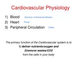
Introduction • The cardiovascular system (CVS) is a closed system in • which the blood circulates throughout the body. It consist of • the heart (pump) and the blood vessels. • Components of the CVS: • Heart: It is a pump composed of 4 chambers (2 atria & 2 • ventricles. • 2. Blood Vessels: The blood vessels are systems of tubes • including: • a) Arteries and arteriols which carry the blood from the • heart to all parts of the body. • b) Venules and veins which carry the blood back from the • tissues to the heart. • c) Blood capillaries which form a network of fine vessels • connecting the arteriols with the venules. The blood • capillaries are the sites of exchange of gases (O2 & CO2), • nutrients and waste products between blood and tissues.
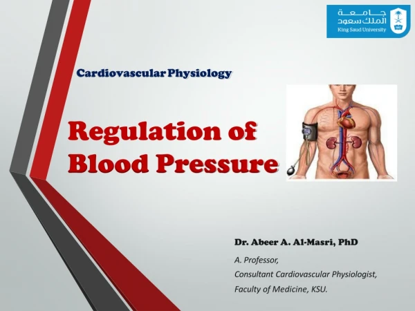
Major Components of the CVS Circulatory System The cardiovascular system (CVS) consists of the heart and blood vessels. It is a closed system in which blood circulates, hence the synonym ‘circulatory system’.
Basic Function of the various parts of the CVS • a) HEART: • 1) The left side of the heart (high pressure side) acts a pressure pump that pumps blood into the systemic arteries at a sufficient pressure that drives blood to the tissues. • 2) The right side of the heart (low pressure side) pumps blood into the pulmonary arteries at a relatively • lower pressure that drives blood into the lungs. • BLOOD VESSELS: • 1) The arteries: the aorta and the pulmonary artery are elastic arteries i.e. they are the properties of stretch (=distension or compliance) and recoil. • During ventricular contraction (systole), they distend by the blood ejected into them; and energy is at load in their walls.
During ventricular relaxation (diastole), this energy is released causing elastic recoil of their walls, which acts as an additional pump to blood during diastole. • Thus on efficient pressure is maintained during systole and diastole, resulting in a continuous blood flow through the tissues. In other words, the arteries act as damping (wind vessel) vessels which convert intermittent pressure into steady pressure that rapidly deliver blood to the tissues. • 2) The arteriols are resistance vessels that act as variable resistors because their diameters continuously undergo changes in order to regulate the amount of blood flow into the capillaries. Therefore, the arterioles are considered the “taps” regulating blood flow to the tissues. • .
3) The capillaries act as exchange vessels between blood and tissues; as through the blood capillaries O2 as nutrients are supplied to the tissues and CO2 as waste products are drained from them. • 4) The veins act as capacitance vessels (volume reservoir) that hold most of the blood volume. Veins have a high distending capacity (=high compliance) and they can store or mobilize blood depending upon the underlying condition. • GENERAL FUNCTION OF THE CVS: • The normal function of the CVS is to maintain homeostasis (i.e. a constant optimum internal environment). Thus, in spite of continuous metabolic activity of the tissue cells, homeostasis is maintained by continuous adequate blood flow to the tissues.
Components: The heart provides the driving force for the cardiovascular system. The arteries serve as distribution channels to the organs. The microcirculation, which includes the capillaries, serve as the exchange region. The veins serve as blood reservoirs and collect the blood to return it to the heart.
The heart The heart is the central pump of the cardiovascular system that drives blood through the blood vessels. It is a muscular structure, which is made up of four chambers. Diastole Systole A heart beat consists of a systole plus a diastole of cardiac chambers. The heart of a normal adult male beats automatically and regularly at a rate of 75 beats/minute during rest. The normal range of heart rate is between 60 – 100. Tachycardia. Bradycardia.
PHYSIOLOGICAL ANATOMY of the HEART • The HEART is the great central pump of the CVS. It lies in the left side of the thoracic cavity partly behind the sternum and between the right and left lungs. It is covered by a fibrous sac called the pericardium. • GENERAL STRUCTURE OF THE HEART • The heart is a hollow muscular organ. Its walls are • composed of a muscle called the cardiac muscle or the • myocardium which is lined by a endothelial layer • called the endocardium (in contact with the blood • inside the heart cavity) and covered by a thin layer • called the epicardium (=visceral layer of the pericardial • sac). • .
Cardiac Chambers & their functions • The humanHEARTis consist of four chambers: • Two atria (right and left) which are separated from each other by the interatrial septum. • Two ventricles (right and left) which are separated from each other by the interventricular septum. The wall of the left ventricle is about 3 times thicker than the wall of the right ventricle. • The ventricular myocardium (wall) is much thicker and stronger than the atrial myocardium (wall). The atrial muscle (of both atria) is completely separated from the ventricular muscle (of both ventricles) by a fibrous ring called AV ring (atrioventricular ring).
The atria have 2 main functions: • 1) They act as blood reservoir for the blood returning back to the heart. • 2) They act as pumps (primer pumps). Atrial • contraction pushes about 25% of the blood filling the ventricles during ventricular diastole and about 75% of the blood that ventricles during their diastole pass passively i.e. by its own weight. • The ventricles, on the other hand,a re the powerful cardiac pumps filling the arteries with blood. The right ventricle (pulmonary pumps) pushes blood into the pulmonary arteries and the left ventricle (systemic pump) pushes blood into the aorta during ventricular systole. • .
CardiacValves and their functions • The human heart contains four valves • Two atrioventricular valves (AV valves) between the • atria and the ventricles: • - Tricuspid valve between the right atrium and the right ventricle. • - Mitral or tricuspid valve between the left atrium and there left ventricle. • Two semilunar valves: • - Aortic valve between the left ventricle and the aorta. • - Pulmonary valve between the right ventricle and the pulmonary trunk. • .
Functions of the cardiac valves • The cardiac valves allow for the blood to pass only in one direction i.e. • - The AV valves allow for the blood to pass from the atria into the ventricles during ventricular diastole. During ventricular systole, the AV valves close to prevent back flow of blood from the ventricles into the atria. • - The semilunar valves allow for the blood to pass from the ventricles into the arteries during ventricular systole. During ventricular diastole, these valves prevent back flow of blood from the arteries into the ventricles (as these valves become closed during ventricular diastole).
It should be noted that: • a) The valves open or close depending upon the pressure gradient of the blood on both sides of the valves e.g. • The AV valves: • - Open when the atrial pressure becomes higher than the ventricular pressure or • - Close when the ventricular pressure becomes higher that the atrial pressure. • The semilunar valves: • - Open when the ventricular pressure becomes higher than the arterial pressure and • - Close when the arterial pressure becomes higher than the ventricular pressure.
Cardiac muscle as a functional sancytum • The cardiac muscle is formed of a network of branching muscle fibers. Each muscle fiber has a separate cell membrane. The sites of contact (end to end contact) between the different fibers are called intercalated discs. Also, the membranes of the adjacent fibers fuse for considerable distances producing gap functions. • These functions provide low electrical resistance for rapid spread of the excitation wave (=cardiac action potential) from one fiber to the other ones. Thus, the cardiac muscle acts as functional sanctum and it contracts as one unit.
The human heart contains 2 separate syncytia, atrial • sancytum (= wall of the two atria) and ventricular • sancytum (= wall of the two ventricles). The two • syncytia are completely separated by a fibrous ring • called the AV ring which prevents passage of the • excitation waves from one sancytum to the other one. • Therefore, there is a special conducting system for • transmission of the excitation waves from the atrial • sancytum to the ventricular sancytum.
The heart as a pump: The heart is two pumps in series (i.e., the right and left sides) that are connected by the pulmonary and systemic circulations.
Systemic Circulation & Pulmonary Circulation In the human body, the blood circulates within the CVS in 2 circulations: • The 1st one to the lungs and it is called the pulmonary or lesser circulation • 1) Systemic or greater circulation: • It starts from the left ventricle and it ends in the right atrium of the heart as follows: • The blood is pumped from the left ventricle of the heart to the aorta, then nutrients are given to the tissue cells and CO2 and waste products of metabolism are taken from them). • The blood is collected from the capillaries by the venules to the veins to the superior and inferior vena cava which carry the blood to the right atrium of the heart.
2) Pulmonary or lesser circulation: • It starts from the right ventricle and it ends in the left atrium of the heart as follows: • The blood is pumped from the right ventricle to the pulmonary arteries & to the pulmonary capillaries (where exchange of O2 and CO2 occurs between the blood and the air in the alveoli of the lungs. • The blood is then collected by the pulmonary veins which carry the blood to the left atrium of the heart. The blood then passes from the LA to LV where systemic circ. begin.
In the cardiovascular system, blood passes through two circulations in series. One full circulation consists of these two circulations together. Both circulations start and end in the heart. These two circulations are: The systemic (or greater or high-pressure) circulation: It starts in the left ventricle → the aorta → systemic arteries → systemic capillaries → systemic veins → superior and inferior vena cavae → ends in the right atrium. The pulmonary (or lesser or low-pressure) circulation: It starts in the right ventricle → the pulmonary trunk → pulmonary arteries → pulmonary capillaries → pulmonary veins → ends in the left atrium.
* The two circulations are in series. So, blood finishes one circulation to start the other. * This allows the whole blood volume to carry out its respiratory function more efficiently as blood goes once through the systemic capillaries and once through the pulmonary capillaries. * Thus, both ventricles must pump the same volume of blood during any significant time interval because of the series arrangement of the systemic and pulmonary circulations. In other words, pulmonary blood flow (right-sided output) must equal systemic blood flow (left-sided output). The balanced output is achieved by an intrinsic property of cardiac muscle known as the Frank-Starling mechanism.
Parallel distribution to the organs * Although the right and left sides of the heart are connected in series, the various systemic organs receive blood flow through parallel distribution channels. * This parallel arrangement supplies systemic organs with blood that has the same arterial composition (e.g., the same O2 and CO2 tensions, pH, glucose levels, and so on) and essentially the same arterial pressure.
Organization in the Circulatory System SERIES AND PARALLEL CIRCUITS
The right ventricle * The right ventricle pumps relatively large volumes of blood at a low pressure through the pulmonary circulation (the right ventricle is essentially flow generator). * The normal cross-section of the right ventricle is crescent-shaped. * If the right ventricle must eject blood against a high pressure for prolonged periods (as seen in certain pulmonary diseases), it assumes a much more cylindrical appearance and there is a thickening of the right ventricular free wall (right ventricular hypertrophy).
The left ventricle The left ventricle pumps blood through the systemic circulation. It is cylindrical in shape and normally has a thicker wall than does the right ventricle. The left ventricle works much harder than the right ventricle because of the higher pressure in the systemic circulation (the left ventricle is essentially pressure generator). Consequently, the left ventricle is more commonly affected by disease processes than is the right ventricle.
Blood flow from the heart During ventricular systole, blood is pumped into the circulation. During diastole, the pumping of blood stops and the ventricles get filled with blood. In this way, the flow of blood from the ventricles into the systemic and pulmonary circulations is an intermittent pulsatile flow.
Cardiac output * Cardiac output is the blood flow generated by each ventricle per minute. * The cardiac output is equal; to the volume of blood pumped by one ventricle per beat times the number of beats per minute: Q = SV . HR Where Q = cardiac output, SV = stroke volume, and HR = heart rate.
The stroke volume for each ventricle averages 70 ml of blood, and a normal heart rate is approximately 70-75 beats/minute; therefore, the cardiac output at rest is approximately 5 L/min. The heart rate is under neural control. Cardiac sympathetic efferent activity increases the heart rate, whereas parasympathetic (vagal) efferent impulses decreases heart rate. The stroke volume varies with the volume of blood in the ventricle at the onset of contraction, changes in the force of ventricular contraction, and the arterial pressure.
Blood vessels 1. Elastic vessels. 2. Low-resistance vessels. 3. High-resistance vessels. 4. Exchange vessels. 5. Capacitance vessels.
Blood pressure What drives blood along the blood vessels after it has left the heart?
The peripheral resistance As the blood flows from the arterial to the venous side of the circulation, it meets resistance because of the smaller caliber of the vessels and the viscous nature of the blood. This is called the peripheral resistance. It is an important factor in generating and maintaining the arterial blood pressure. Vasoconstriction of the small vessels increases the peripheral resistance, which in turn elevates the arterial blood pressure. Whilst vasodilatation decreases the resistance and lowers the pressure. The main factor is a gradient of blood pressure.
RESISTANCES IN SERIES RT = RA + RC + RV RESISTANCES IN PARALLEL R1 PV PA 1 RT 1 R1 1 R2 1 R3 R2 = + + R3 1 RT = 1 R1 1 R2 1 R3 + +
Pressure Drop in the Vascular System ELASTIC TISSUE MUSCLE LARGEARTERIES SMALLARTERIES MEANPRESSURE ARTERIOLES CAPILLARIES VENULES &VEINS LARGE SMALL LARGE INSIDE DIAMETER