Download

1 / 76
820 likes | 1.64k Vues
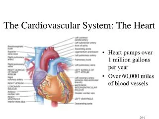
The Cardiovascular System: The Heart Anatomy. 18. Heart Anatomy. Approximately the size of your fist Location Superior surface of diaphragm Left of the midline Anterior to the vertebral column, posterior to the sternum. Heart Anatomy. Figure 18.1. Coverings of the Heart: Anatomy.

E N D
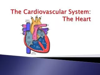
The Cardiovascular System: The Heart Anatomy 18 Chapter 18, Cardiovascular System
Heart Anatomy • Approximately the size of your fist • Location • Superior surface of diaphragm • Left of the midline • Anterior to the vertebral column, posterior to the sternum Chapter 18, Cardiovascular System
Heart Anatomy Figure 18.1 Chapter 18, Cardiovascular System
Coverings of the Heart: Anatomy • Pericardium – a double-walled sac around the heart composed of: • A superficial fibrous pericardium • A deep two-layer serous pericardium • The parietal layer lines the internal surface of the fibrous pericardium • The visceral layer or epicardium lines the surface of the heart • They are separated by the fluid-filled pericardial cavity Chapter 18, Cardiovascular System
Coverings of the Heart: Physiology • The Function of the Pericardium: • Protects and anchors the heart • Prevents overfilling of the heart with blood • Allows for the heart to work in a relatively friction-free environment Chapter 18, Cardiovascular System
Pericardial Layers of the Heart Figure 18.2 Chapter 18, Cardiovascular System
Heart Wall • Epicardium – visceral layer of the serous pericardium • Myocardium – cardiac muscle layer forming the bulk of the heart • Fibrous skeleton of the heart – crisscrossing, interlacing layer of connective tissue • Endocardium – endothelial layer of the inner myocardial surface Chapter 18, Cardiovascular System
External Heart: Major Vessels of the Heart (Anterior View) • Vessels returning blood to the heart include: • Superior and inferior venae cavae • Right and left pulmonary veins • Vessels conveying blood away from the heart include: • Pulmonary trunk, which splits into right and left pulmonary arteries • Ascending aorta (three branches) – • Brachiocephalic • Left common carotid • Subclavian arteries Chapter 18, Cardiovascular System
External Heart: Vessels that Supply/Drain the Heart (Anterior View) • Arteries – right and left coronary (in atrioventricular groove), marginal, circumflex, and anterior interventricular arteries • Veins – small cardiac, anterior cardiac, and great cardiac veins Chapter 18, Cardiovascular System
External Heart: Anterior View Figure 18.4b Chapter 18, Cardiovascular System
External Heart: Major Vessels of the Heart (Posterior View) • Vessels returning blood to the heart include: • Right and left pulmonary veins • Superior and inferior venae cavae • Vessels conveying blood away from the heart include: • Aorta • Right and left pulmonary arteries Chapter 18, Cardiovascular System
External Heart: Vessels that Supply/Drain the Heart (Posterior View) • Arteries – right coronary artery (in atrioventricular groove) and the posterior interventricular artery (in interventricular groove) • Veins – great cardiac vein, posterior vein to left ventricle, coronary sinus, and middle cardiac vein Chapter 18, Cardiovascular System
External Heart: Posterior View Chapter 18, Cardiovascular System Figure 18.4d
Gross Anatomy of Heart: Frontal Section Figure 18.4e Chapter 18, Cardiovascular System
Atria of the Heart • Atria are the receiving chambers of the heart • Each atrium has a protruding auricle • Pectinate musclesmark atrial walls • Blood enters right atria from superior and inferior venae cavae and coronary sinus • Blood enters left atria frompulmonary veins Chapter 18, Cardiovascular System
Ventricles of the Heart • Ventricles are the discharging chambers of the heart • Papillary muscles and trabeculae carneae muscles mark ventricular walls • Right ventriclepumps blood into the pulmonary trunk • Left ventriclepumps blood into the aorta Chapter 18, Cardiovascular System
Myocardial Thickness and Function Thickness of myocardium varies according to the function of the chamber Atria are thin walled, deliver blood to adjacent ventricles Ventricle walls are much thicker and stronger • right ventricle supplies blood to the lungs (little flow resistance) • left ventricle wall is the thickest to supply systemic circulation Chapter 18, Cardiovascular System
Thickness of Cardiac Walls Myocardium of left ventricle is much thicker than the right. Chapter 18, Cardiovascular System
Atrial Septal Defect Chapter 18, Cardiovascular System
Ventricular Septal Defect Chapter 18, Cardiovascular System
Pathway of Blood Through the Heart and Lungs • Right atrium tricuspid valve right ventricle • Right ventricle pulmonary semilunar valve pulmonary arteries lungs • Lungs pulmonary veins left atrium • Left atrium bicuspid valve left ventricle • Left ventricle aortic semilunar valve aorta • Aorta systemic circulation Chapter 18, Cardiovascular System
Pathway of Blood Through the Heart and Lungs Figure 18.5 Chapter 18, Cardiovascular System
Coronary Circulation • Coronary circulation is the functional blood supply to the heart muscle itself • Collateral routesensure blood delivery to heart even if major vessels are occluded Chapter 18, Cardiovascular System
Coronary Circulation: Arterial Supply Chapter 18, Cardiovascular System Figure 18.7a
Coronary Circulation: Venous Supply Chapter 18, Cardiovascular System Figure 18.7b
Heart Valves • Heart valves ensure unidirectional blood flow through the heart • Atrioventricular (AV) valves lie between the atria and the ventricles • AV valves prevent backflow into the atria when ventricles contract • Chordae tendineae anchor AV valves to papillary muscles Chapter 18, Cardiovascular System
Heart Valves • Semilunar valves prevent backflow of blood into the ventricles • Aortic semilunar valve lies between the left ventricle and the aorta • Pulmonary semilunar valve lies between the right ventricle and pulmonary trunk Chapter 18, Cardiovascular System
Heart Valves Figure 18.8a, b Chapter 18, Cardiovascular System
Heart Valves Figure 18.8c, d Chapter 18, Cardiovascular System
Atrioventricular Valve Function Figure 18.9 Chapter 18, Cardiovascular System
Semilunar Valve Function Figure 18.10 Chapter 18, Cardiovascular System
Mitral Valve Prolapse Chapter 18, Cardiovascular System
Microscopic Anatomy of Heart Muscle • Cardiac muscle is striated, short, fat, branched, and interconnected • The connective tissue endomysium acts as both tendon and insertion • Intercalated discs anchor cardiac cells together and allow free passage of ions • Heart muscle behaves as a functional syncytium InterActive Physiology®: Cardiovascular System: Anatomy Review: The Heart PLAY Chapter 18, Cardiovascular System
Microscopic Anatomy of Heart Muscle Chapter 18, Cardiovascular System Figure 18.11
The Cardiovascular System: The Heart Physiology 18 Chapter 18, Cardiovascular System
Cardiac Muscle Contraction • Heart muscle: • Is stimulated by nerves and is self-excitable (automaticity) • Contracts as a unit • Has a long (250 ms) absolute refractory period • Cardiac muscle contraction is similar to skeletal muscle contraction Chapter 18, Cardiovascular System
Heart Physiology: Intrinsic Conduction System • Autorhythmic cells: • Initiate action potentials • Have unstable resting potentials called pacemaker potentials • Use calcium influx (rather than sodium) for rising phase of the action potential Chapter 18, Cardiovascular System
Pacemaker and Action Potentials of the Heart Chapter 18, Cardiovascular System Figure 18.13
Heart Physiology: Sequence of Excitation • Sinoatrial (SA) node generates impulses about 75 times/minute • Atrioventricular (AV) node delays the impulse approximately 0.1 second Chapter 18, Cardiovascular System
Heart Physiology: Sequence of Excitation • Impulse passes from atria to ventricles via the atrioventricular bundle (bundle of His) • AV bundle splits into two pathways in the interventricular septum (bundle branches) • Bundle branches carry the impulse toward the apex of the heart • Purkinje fibers carry the impulse to the heart apex and ventricular walls Chapter 18, Cardiovascular System
Heart Physiology: Sequence of Excitation Chapter 18, Cardiovascular System Figure 18.14a
Heart Excitation Related to ECG Figure 18.17 Chapter 18, Cardiovascular System
Extrinsic Innervation of the Heart • Heart is stimulated by the sympathetic cardioacceleratory center • Heart is inhibited by the parasympathetic cardioinhibitory center Chapter 18, Cardiovascular System Figure 18.15
Electrocardiography • Electrical activity is recorded by electrocardiogram (ECG) • P wave corresponds to depolarization of SA node • QRS complex corresponds to ventricular depolarization • T wave corresponds to ventricular repolarization • Atrial repolarization record is masked by the larger QRS complex InterActive Physiology®: Cardiovascular System: Intrinsic Conduction System PLAY Chapter 18, Cardiovascular System
Electrocardiography Figure 18.16 Chapter 18, Cardiovascular System
Heart Sounds • Heart sounds (lub-dup) are associated with closing of heart valves • First sound occurs as AV valves close and signifies beginning of systole (contraction) • Second sound occurs when SL valves close at the beginning of ventricular diastole (relaxation) Chapter 18, Cardiovascular System
Cardiac Cycle • Cardiac cycle refers to all events associated with blood flow through the heart • Systole – contraction of heart muscle • Diastole – relaxation of heart muscle Chapter 18, Cardiovascular System
Phases of the Cardiac Cycle • Ventricular filling – mid-to-late diastole • Heart blood pressure is low as blood enters atria (passively) and flows into ventricles • AV valves are open, then atrial systole occurs Chapter 18, Cardiovascular System
Phases of the Cardiac Cycle • Ventricular systole (contraction) • Atria relax • Rising ventricular pressure results in closing of AV valves • Isovolumetric contraction phase • Ventricular ejection phase opens semilunar valves Chapter 18, Cardiovascular System
Phases of the Cardiac Cycle • Isovolumetric relaxation – early diastole • Ventricles relax • Backflow of blood in aorta and pulmonary trunkcloses semilunar valves • Dicrotic notch – brief rise in aortic pressure caused by backflow of bloodrebounding off semilunar valves InterActive Physiology®: Cardiovascular System: Cardiac Cycle PLAY Chapter 18, Cardiovascular System