The Cardiovascular System: The Heart
600 likes | 848 Vues
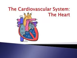
The Cardiovascular System: The Heart. Heart Anatomy. Approximately the size of your fist Location: • Superior surface of diaphragm • Left of the midline • Anterior to the vertebral column, posterior to the sternum. Heart Covering. Pericardial physiology Protects and anchors heart

The Cardiovascular System: The Heart
E N D
Presentation Transcript
Heart Anatomy • Approximately the size of your fist Location: • Superior surface of diaphragm • Left of the midline • Anterior to the vertebral column, posterior to the sternum
HeartCovering • Pericardial physiology • Protects and anchors heart • Prevents overfilling • Pericardial anatomy • Fibrous pericardium • Serous pericardium (separated by pericardial cavity) • Epicardium (visceral layer)
Heart Wall • Epicardium • visceral layer of the serous pericardium • Myocardium • cardiac muscle layer forming the bulk of the heart • Fibrous skeleton of the heart • crisscrossing, interlacing layer of connective tissue • Endocardium • endothelial layer of the inner myocardial surface
ExternalHeart: Major Vessels of the Heart (Anterior View) • Returning blood to the heart • Superior and inferior venae cavae • Right and left pulmonary veins • Conveying blood away from the heart • Pulmonary trunk, which splits into right and left pulmonary arteries • Ascending aorta (three branches) • brachiocephalic, left common carotid, and subclavian arteries
External Heart: Vessels that Supply/Drain the Heart (Anterior View) • Arteries • right and left coronary (in atrioventricular groove), marginal, circumflex, and anterior interventricular • Veins • small cardiac vein, anterior cardiac vein, and great cardiac vein
External Heart: Major Vessels of the Heart (Posterior View) • Returning blood to the heart • Right and left pulmonary veins • Superior and inferior venae cavae • Conveying blood away from the heart • Aorta • Right and left pulmonary arteries
External Heart: Vessels that Supply/Drain the Heart (Posterior View) • Arteries • coronary artery (in atrioventricular groove) and the posterior interventricular artery (in interventricular groove) • Veins • great cardiac vein, posterior vein to left ventricle, coronary sinus, and middle cardiac vein
Gross Anatomy of Heart: Frontal Section • Frontal section showing interior chambers and valves • Major vessels leading to and from the heart
Gross Anatomy of Heart: Frontal Section Atria of the Heart • Atria are the receiving chambers of the heart • Each atrium has a protruding auricle • Pectinate muscles mark atrial walls • Blood enters right atria from superior and inferior venaecavae and coronary sinus • Blood enters left atria from pulmonary veins
Ventricles of the Heart • Ventricles are the discharging chambers of the heart • Papillary muscles and trabeculae carneae muscles mark ventricular walls • Right ventricle pumps blood into the pulmonary trunk • Left ventricle pumps blood into the aorta
Pathway of Blood through the Heart and Lungs • Right atrium tricuspid valve right ventricle • Right ventricle pulmonary semilunar valve pulmonary arteries lungs • Lungs pulmonary veins left atrium • Left atrium bicuspid valve left ventricle • Left ventricle aortic semilunar valve aorta • Aorta systemic circulation
Coronary Circulation • Coronary circulation is the functional blood supply to the heart • Collateral routes insure blood delivery to heart even if major vessels are occluded
Heart Valves • Heart valves insure unidirectional blood flow through the heart • Atrioventricular (AV) valves lie between the atria and the ventricles • AV valves prevent backflow into the atria when ventricles contract • Chordaetendineae anchor AV valves to papillary muscles • Aortic semilunar valve lies between the left ventricle and the aorta • Pulmonary semilunar valve lies between the right ventricle and pulmonary trunk • Semilunar valves prevent backflow of blood into the ventricles
Microscopic Heart Muscle Anatomy • Cardiac muscle is striated, short, fat, branched, and interconnected • Connective tissue endomysium acts as both tendon and insertion • Intercalated discs anchor cardiac cells together and allow free passage of ions • Heart muscle behaves as a functional syncytium
Cardiac Muscle Contraction • Heart muscle: • Is stimulated by nerves and self-excitable (automaticity) • Contracts as a unit • Has a long (250 ms) absolute refractory period • Cardiac muscle contraction is similar to skeletal muscle contraction
Heart Physiology:Intrinsic Conduction System • Autorhythmic cells: • Initiate action potentials • Have unstable resting potentials called pacemaker potentials • Use calcium influx (rather than sodium) for rising phase of the action potential
Heart Physiology: Sequence of Excitation • Sinoatrial (SA) node generates impulses about 75 times/minute • Atrioventricular (AV) node delays the impulse approximately 0.1 second • Impulse passes from atria to ventricles via the atrioventricular bundle (bundle of His)
Heart Physiology: Sequence of Excitation • AV bundle splits into two pathways in the interventricular septum (bundle branches) • Bundle branches carry the impulse toward the apex of the heart • Purkinje fibers carry the impulse to the heart apex and ventricular walls
Extrinsic Innervation of the Heart • Heart is stimulated by the sympathetic cardioacceleratory center • Heart is inhibited by the parasympathetic cardioinhibitory center
Electrocardiography • Electrical activity is recorded by electrocardiogram (ECG) • P wave corresponds to depolarization of SA node • QRS complex corresponds to ventricular depolarization • T wave corresponds to ventricular repolarization • Atrialrepolarization record is masked by the larger QRS complex
Cardiac Cycle • Cardiac cycle refers to all events associated with blood flow through the heart • Systole – contraction of heart muscle • Diastole – relaxation of heart muscle
Phases of the Cardiac Cycle • Ventricular filling – mid-to-late diastole • Heart blood pressure is low as blood enters atria and flows into ventricles • AV valves are open then atrial systole occurs • Ventricular systole • Atria relax • Rising ventricular pressure results in closing of AV valves • Isovolumetric contraction phase • Ventricular ejection phase opens semilunar valves
Phases of the Cardiac Cycle • Isovolumetric relaxation - early diastole • Ventricles relax • Backflow of blood in aorta and pulmonary trunk closes semilunar valves • Dicrotic notch – brief rise in aortic pressure caused by backflow of blood rebounding off semilunar valves
Heart Sounds • Heart sounds (lub-dup) are associated with closing of heart valves
Cardiac Output (CO) and Reserve • CO is the amount of blood pumped by each ventricle in one minute • CO is the product of heart rate (HR) and stroke volume (SV) • HR is the number of heart beats per minute • SV is the amount of blood pumped out by a ventricle with each beat • Cardiac reserve is the difference between resting and maximal CO
Cardiac Output: Example • CO (ml/min) = HR (75 beats/min) x SV (70 ml/beat) • CO = 5250 ml/min (5.25 L/min)
Regulation of Stroke Volume • SV = end diastolic volume (EDV) minus end systolic volume (ESV) • EDV = amount of blood collected in a ventricle during diastole • ESV = amount of blood remaining in a ventricle after contraction
Factors Affecting Stroke Volume • Preload – amount ventricles are stretched by contained blood • Contractility – cardiac cell contractile force due to factors other than EDV • Afterload – back pressure exerted by blood in the large arteries leaving the heart
Frank-Starling Law of the Heart • Preload, or degree of stretch, of cardiac muscle cells before they contract is the critical factor controlling stroke volume • Slow heartbeat and exercise increase venous return to the heart, increasing SV • Blood loss and extremely rapid heartbeat decrease SV
Preload and AfterloadExtrinsic Factors Influencing Stroke Volume • Contractility is the increase in contractile strength, independent of stretch and EDV • Increase in contractility comes from: • Increased sympathetic stimuli • Certain hormones • Ca2+ and some drugs • Agents/factors that decrease contractility include: • Acidosis • Increased extracellular potassium • Calcium channel blockers
Contractility and Norepinephrine • Sympathetic stimulation releases norepinephrine and initiates a cyclic AMP second-messenger system
Regulation of Heart Rate: Autonomic Nervous System • Sympathetic nervous system (SNS) stimulation is activated by stress, anxiety, excitement, or exercise • Parasympathetic nervous system (PNS) stimulation is mediated by acetylcholine and opposes the SNS • PNS dominates the autonomic stimulation, slowing heart rate and causing vagal tone
Bainbridge Reflex • Bainbridge (atrial) reflex – a sympathetic reflex initiated by increased blood in the atria • Causes stimulation of the SA node • Stimulates baroreceptors in the atria, causing increased SNS stimulation
Chemical Regulation of the Heart • The hormones epinephrine and thyroxine increase heart rate • Intra- and extracellular ion concentrations must be maintained for normal heart function
Factors Involved in Regulation of Cardiac OutputHomeostatic Imbalances • Hypocalcemia • reduced ionic calcium depresses the heart • Hypercalcemia • dramatically increases heart irritability and leads to spastic contractions • Hypernatremia • blocks heart contraction by inhibiting ionic calcium transport • Hyperkalemia • leads to heart block and cardiac arrest
Homeostatic Imbalances • Tachycardia – heart rate over 100 beats/min • Bradycardia – heart rate less than 60 beats/min