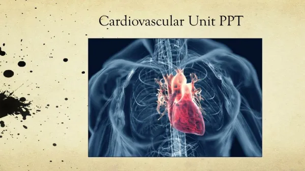
Cardiovascular Unit
Cardiovascular Unit. By Pat Pence, RN, C, MSN. Heart. Located in mediastinum. Base (wider) is superior and under the 2 nd rib. Apex (narrow) is inferior and slightly to the left between 5 th and 6 th ribs. . Heart wall. Composed of 3 layers.

Cardiovascular Unit
E N D
Presentation Transcript
Cardiovascular Unit By Pat Pence, RN, C, MSN
Heart • Located in mediastinum. • Base (wider) is superior and under the 2nd rib. • Apex (narrow) is inferior and slightly to the left between 5th and 6th ribs.
Heart wall • Composed of 3 layers. • Pericardium: transparent, thin layer, lines outside of heart, 2 layers that contain serous fluid to decrease friction. • Myocardium: middle, thickest and strongest, actual contracting muscle tissue. • Endocardium: innermost layer, thin layer of connective tissue.
4 Heart chambers • Right atrium: receives deoxygenated blood from superior vena cava, inferior vena cava and coronary sinus. • Right ventricle:pumps blood to lungs via pulmonary artery. • Left atrium: receives O2 rich blood from lungs via pulmonary veins. • Left ventricle: PMI, thickest, most muscular, pumps blood to all parts of body via aorta. • Separated by septum.
4 Heart valves • Heart functions as 2 separate pumps. • Heart valves keep blood flowing forward and prevent backflow (regurgitation). • Tricuspid valve and mitral valve (AV valves). • Chordae tendineae and papillary muscles connect valves to walls of heart and promote a tight seal to prevent backflow.
Semilunar valves • Pulmonary semilunar valve: between Rt ventricle and pulmonary artery. • Aortic semilunar valve: between left ventricle and aorta.
Coronary blood supply • Heart requires a constant supply of O2 rich blood and return of O2 poor blood from tissue to the lungs. • Rt./Lt. Coronary arteries. • Coronary vein and coronary sinus. • Collateral circulation.
Blood vessel pattern • Artery: largest, vessels that carry blood away from heart; thicker, elastic and muscle tissue. • Arteriole: smooth muscle, deliver blood to tissues; dilate or constrict in response to low O2/hi CO2, affect BP blood flow. • Capillary: endothelial cells, allow exchange of products; no muscle or elastic tissue.
Venules: small amounts of muscle and connective tissue. • Veins: larger veins have valves to prevent backflow of blood; carry blood back to heart; large diameter, thin walled.
Pulmonary circulation • Deoxygenated blood passes through pulmonary circulation to receive O2. • Right ventricle, pulmonary semilunar valve, pulmonary artery, pulmonary capillaries, pulmonary veins, left atrium, bicuspid valve, left ventricle, aortic semilunar valve, and aorta.
Systemic circulation • Refers to blood pumped from lt ventricle to all parts of body and then to rt atrium. • Aorta: largest artery, main trunk of systemic arterial circulation. • Vena cava: returns deoxygenated blood to rt atrium. • Superior vena cava: from head, neck, chest, and upper extremities. • Inferior vena cava: from parts of body below diaphragm.
Electrical conduction system • Automaticity: specialized ability to contract in rhythmic pattern. • Irritability: excitability or sensitivity. • Hormones, ion concentration, changes in body temp. affect the conduction system. • Depolarization = contraction. • Repolarization = relaxation (resting state).
Impulse pattern • Initiated in SA node in Rt. Atrium. “Pacemaker” of heart. Internodal pathways to atria. • AV node: Rt atrium, slows impulses to allow atrium to contract and ventricles to fill. • Bundle of His: group of conduction fibers in base of rt atrium. • Rt. and Lt. Bundle branches in septum. • Perkinje’s fibers: surround ventricles.
Cardiac cycle: systole/diastole • Refers complete heartbeat. • Systole: = contraction, blood is pumped out of the ventricles. (depolarization). • Atria relax to receive blood. (repolarization). • AV valves close: S1: first heart sound. • Ventricles contract in response to electrical impulse having passed through.
Diastole • Diastole = relaxation, blood enters the relaxed chambers. • Ventricles fill. • Semilunar valves close: S2: 2nd heart sound. • Atria contract in response to electrical impulse.
Stroke volume/cardiac output • Stroke volume: volume of blood ejected (pumped) during each ventricular contraction (heartbeat). • Cardiac output: amount of blood ejected (pumped) by each ventricle per minute. • CO = SV X HR. • Numerous factors affect CO.
Stroke volume factors • Preload: volume of blood within ventricles at end of diastole, comes from veins, before next contraction. Preload determines the amount of stretch placed on the myocardium. • Afterload: pressure that ventricles work against when they contract to eject blood from the heart. That pressure is located in arteries (peripheral resistance or arterial BP).
Normal heart overcomes afterload and maintains CO except when muscles have been damaged. • Contractility increased by norepinephrine and epinephrine. • Increased preload, afterload and contractility increase workload and O2 demand on heart.
Inotropic state • Inotropic state = strength of myocardial contraction unrelated to blood volume. • Affected by sympathetic stimulation, metabolic abnormality, hypoxemia, metabolic acidosis, drugs (epinephrine).
Starling’s law • To a point the heart will pump out all blood it receives within certain limits. • The more the fibers are stretched, the greater their force of contraction. • If stretched beyond capacity- blood will accumulate in ventricles and back up into pulmonary system.
Heart sounds • Produced by closure of valves. • Lubb: (S1) long duration, low pitch, AV valves close. • Dubb: (S2) short duration, sharp sound, semilunar valves close. • Murmur: swishing sound, may be normal or abnormal.
Cardiovascular assessment • Subjective data: past CV problems, health habits (smoking, diet, activity), current CV problems. • History: description of symptoms, when they occurred, course and duration, location, precipitating factors, relief measures.
Pain • Character, quality, radiation, associated symptoms. • Rated on pain scale. • Location: *chest, radiated to jaw, left shoulder. • Description: dull, sharp, pressure, squeezing, crushing, viselike, grinding, radiating. • Precipitating onset. • Pain in extremities or lack of sensation.
Palpitations • Characterized by rapid, irregular, or pounding heartbeat. • Associated with dysrhythmias or ischemia.
Dyspnea • Exertional dyspnea is associated with decreased cardiac output. • DOE, DAR, PND, orthopnea.
Cough • Dry, productive, irritating, spasmodic. • May be associated with dyspnea.
Fatigue • Exhaustion/ activity intolerance. • Associated with decreased cardiac output. • Depression may associated with fatigue.
Syncope • Fainting: brief lapse of consciousness. • Caused by transient cerebral hypoxia. • Sudden decrease in cardiac output to brain resulting from dysrhythmia or decreased pumping action of heart. • Preceded by lightheadedness.
Objective data • Vital signs. LOC. • Lung sounds. Bowel sounds. Sputum. • Heart: Apical pulse, heart sounds, carotid arteries, systemic with no bruit. • Jugular veins not distended when sitting or standing. • Extremities: color, temp, moisture, edema, hair distribution, capillary refill, clubbing, peripheral pulses, Homan’s sign, turgor.
Cyanosis • Caused by: excess of deoxygenated Hgb in blood. • Results from: decreased cardiac output and poor peripheral perfusion.
Diaphoresis • Profuse sweating associated with clamminess. • Result of decreased cardiac output and poor peripheral perfusion.
Edema • Wt. Gain of more than 3lb. In 24 hours. • Inability of heart to pump efficiently or accept venous return, causes backflow of blood and increased blood volume. • Increased hydrostatic pressure results and an increase in fluid to interstitial spaces.
CBC • Determination of RBCs, WBCs, platelets, Hgb, Hct. • Low Hgb = decreased O2 carrying capacity • High WBC = infection or inflammation. • High RBC = body compensating for chronic hypoxemia by stimulating RBC production in bone marrow, leading to secondary polycythemia.
Cardiac enzymes • Elevated amounts of enzymes (proteins, cardiac “markers”) are released into blood during cardiac muscle cell damage. • These enzymes are helpful in determining the degree of myocardial damage and the timing of onset of damage. • Some enzymes are more specific to cardiac muscle tissue.
Creatine phosphokinase (CPK) • Enzyme found in brain, skeletal muscle, and myocardium. • Can be broken down into isoenzymes. • CPK II (MB) is more specific to cardiac tissue. Levels > 7.5 ng/ml associated c MI. • Onset: 3-6 hours; peak 12-18 hours; duration 3-4 days.
Lactic dehydrogenase (LDH) • Found in many body tissues (cardiac, kidneys, RBCs, brain, stomach, skeletal muscle). Normal < 100 U/L. • Late indicator of damage. • Rises in 24-72 hours; peaks in 3-4 days; returns to normal levels in about 14 days. • Not as specific as other enzymes.
Myoglobin • Small O2 binding protein found in cardiac and skeletal muscle. • Because it is smaller when compared to larger enzymes, it is detected earlier. • 1-4 hours after onset. Peak 6-9 hrs. • Must be done within 1st 18 hrs after onset. • Normal < 92 men; < 76ng/ml women.
Troponin • Protein located on cardiac and skeletal muscle tissue. • 3 forms. Troponin I is more specific; found exclusively in cardiac tissue. • Onset 3-6 hours; peaks 14-20 hours; returns to normal in 5-7 days. • Elevated levels of Troponin I is diagnostic of MI. Range 0 - 0.4ng/mL.
Serum Lipids • Cholesterol: fatty substance coated with 2 types of proteins. LDL or HDL. • Total cholesterol level is sum of all cholesterols. Level should be < 200mg/dl. (nonfasting test). • Associated with diet high in saturated fat, cholesterol and calories.
Hyperlipidemia: elevated levels of any or all of lipids in plasma. • Triglycerides: Mixture of fatty acids. • Normal: 40-190 ng/dl.
HDL/LDL • HDL and LDL: need to fast 12 hrs. • High-density lipoprotein: “good cholesterol”. More protein than fat. • Removes cholesterol from vessels and transports to liver for removal. • Higher the level = less risk of CAD. • Desirable level = 35 or >.
LDL • Low-density lipoprotein: “bad cholesterol”. Promotes CAD. • Equal amount of fat and protein. • Transports cholesterol from the liver to body tissues, accumulating in vessel walls. • Level < 160mg/dl (c no risk factors). Higher the level = greater risk of CAD. Desired level for LDL depends on the risk factor profile of pt. More risks = a goal for a lower LDL level. (ex. 100mg/dl)
EKG • A graphic representation of the heart’s electrical activity reflected by changes in the electrical potential at the skin surface. • Recorded as a tracing on a strip of paper. • “Resting” EKG over < one minute. • Telemetry: continuous visualization on a screen (monitor).
Purposes • Identify rhythm disturbances. • Provides information about the position of the heart in the chest and the size of the chambers. • Detects electrolyte imbalances. • Monitors the effectiveness of pacemakers and cardiotonic meds.
Holter monitor • “Ambulatory” EKG: a type of portable EKG which records 12- 48 hours of usual pt.’s activities with normal stress. • Pt keeps a diary of activities and symptoms. • Purposes: determines exercise tolerance post-MI. Provides more diagnostic information.
Inpatient or outpatient basis. • Detects intermittent arrythmias c ADLs c associated manifestations of dizziness, palpitations, chest pain. • Guages antiarrythmic meds. • Instruct pt to update diary regularly and note any symptoms; not to get monitor wet.
EKG waveforms • P wave: depolarization or contraction of the atria. SA node fires impulse, delay by AV node seen as space after P wave. • QRS complex: ventricles depolarize or contract, atria repolarize (relax), strong signal because of greater mass of ventricles. • T wave: ventricles repolarize or relax.
Intervals are the length of time it takes the impulse to travel from one area of heart to another. • Cardiac dysrhythmias result of abnormal pacemaker function.