Download

1 / 41
620 likes | 3.44k Vues
Synovial Fluid Analysis. Definition. Alternative names: Joint fluid analysis; Joint fluid aspiration Synovial fluid is viscous colorless liquid that found in the joint cavities. Formation of synovial fluid.

E N D
Definition • Alternative names: Joint fluid analysis; Joint fluid aspiration • Synovial fluid is viscous colorless liquid that found in the joint cavities.
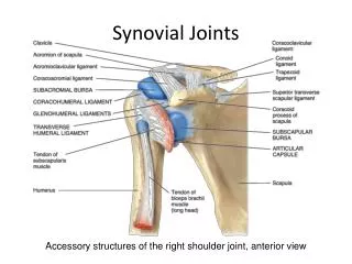
Formation of synovial fluid • Formed as an ultrafiltrate of the plasma across the synovial membrane into which a mucopolysacharide containing hyaluronic acid and small amount of protein (low MWT) is secreted by the cells of synovial membrane. • Except for High MWT proteins (more than 12 kilo Dalton in size), the plasma filtration is non-selective; therefore, normal synovial fluid has essentially the same chemical composition as the plasma.
Function of synovial fluid • Lubricate the joint space; as a lubricant to the surfaces of the frequently moving joints. • Supplies nutrients to particular cartilage; as nutrition must be provided by moving synovial fluid in and out of the cartilage, it may be clear that joint movement is essential to cartilage nutrition and maintenance. • Shock absorber; our cartilage, immersed in the synovial fluid, protects our bones from the tremendous impact they would receive when we walk, run, jump, etc. This fluid also has remarkable properties as a shock absorbing, or hydraulic fluid
Why the test is performed ? • To diagnose certain types of arthritis and inflammatory joint diseases, and relieve pain and distention from fluid the cause of swelling in the accumulation in the joint.
Types of joints disorders 1. Non-inflammatory • Degenerative joint disorders 2. Inflammatory • Rheumatoid arthritis • Lupus erythematosus • Rheumatic fever • Crystal-induced gout • 3. Septic Microbial infection • 4. Hemorrhagic • Traumatic injury • Tumors • Hemophilia and other coagulation disorders
Sample Collection (Arthrocentesis ) • The Fluid is collected by a physician by means of needle aspiration of the knee called arthrocentesis. • Arthrocentesis (removal of SF from a joint) done when patient presents with effusion • The normal amount of fluid contained in the knee cavity is less than 3.5 ml and increased in joint disorders.
Fluid from syringe is divided into 3 tubes: • 2-5 ml in EDTA tube for cell count, crystal exam • 5-10 ml in plain sterile tube for microbiology (culture and stain). • Few ml in Plain tube for Chem. and Immunologic tests. • Liquid EDTA or Na heparin may be used while powdered EDTA and lithium heparin should be avoided as anticoagulant).
Total volume • The total volume collected is recorded on each time. Viscosity • Normal synovial fluid is highly viscous due to the polymerization of the hyaluronic acid, which is essential for the proper lubrication of joints
Joint fluid is aspirated into a pipette and then released • If the falling drop is drawn out into a 4 to 6 cm long or longer tenacious band the viscosity is normal .If the drop falls like water, the viscosity is low.
Decrease synovial fluid viscosity 1. Depolymerization of hyaluronate complex which present in the following conditions: • Rheumatoid arthritis. • septic arthritis. • gout. 2. Dilution of hyaluronate complex or even decrease production due to rabid effusion which take place in trauma
The mucin clot formation test Principle: • Mucin (a hyaluronic acid –protein complex) is precipitated by acetic acid the morphology of the precipitated is a reflection of the hyaluronic acid content and quality of the joint fluid. Reagent: • Acetic acid, 7N: Mix 408 ml glacial acetic acid and 1 L distilled water.
Procedure: 1. Add 1 ml joint fluid to 4 ml distilled water in a test tube. 2. Add 0.14 ml of 7N Acetic acid and stir briskly with a glass rod. 3. Examine immediately and after 2 hr. Interpretation: If a hyaluronic acid is normal, a tight ropy mass forms in a clear solution, indicating “good “ mucin. “Fair” mucin is indicated by a softer in a turbid solution precipitated that shreds into the solution. ”poor” precipitated consists of shreds
Clear fluid; solid clot Friable clot
Clot formation • Because of lack of fibrinogen and other clotting factors, normal joint fluid not clots. • Inflammatory processes allow the plasma clotting factors to escape into the joint fluid, which then clot.
Color and Transparency • Color : Normal SF is pale yellow • Abnormal colors: • 1. Dark red or dark brown (bloody) {hemarthrosis} due to: • Fracture through the joint surface • Tumor involving the joint • Traumatic arthritis • Hemophilic arthritis • May present in septic or rheumatoid arthritis.
2. Deeper yellow or Green tinge: • Bacterial infection. • Chronic rheumatoid arthritis. 3. Xanthochromiawhich is difficult to be evaluated owing to the yellow color of synovial fluid, hence any color is usually due to hemarthrosis. 4. Milky : • Gouty arthritis • Tuberculousarthritis • Calcium hydroxyapatite crystals
2. Transparency: Normal = crystal clear • Turbid : leukocytosis (more than 200 cell/µl). • Cloudy Milky, fatty: • Cholesterol crystals. • Fibrin. • Degenerative synovial cells which gives free floating tissue aggregates.
Cell count & Differential Reagent and procedure • The white cell counting technique is used, but isotonic saline solution (ex. 0.3 saline, 0.1 HCL, 1% saponin in saline) is substituted the usual acetic acid since the latter precipitates the hyaluronic acid –protein complex and that lead to WBCs clumping due to mucin clot. • The cell count should be completed without delay to prevent spontaneous clumping of leukocytes. .
Degeneration of leukocytes begin one hour after sample collection • If the fluid is highly viscous it must be incubate at 37 C with hyaluronidase enzyme 0.05% for 5 min. • Normal value: 200 WBCs/µl. • RBCs usually present in very low numbers but may be present because of trauma of aspiration
Differential cells count: • Mononuclear cells including Monocytes, lymphocytes, macrophage and synovial tissue cells are the primary cells in the normal synovial fluid • Polymorphnuclear cells 0-25% (mean=6%) • Lymphocytes 0-78% (mean=25%) • Monocytes 0-71% (mean=48%) • Macrophages 0-26% (mean=10%)
Wet preparation : • No crystals • No rheumatoid arthritis cells • No cartilage fibers • No bacteria Crystals that may be found: • Crystals indicate the presence of crystal induced arthritis. • Test must be performed at the collection time because crystals are affected by pH, temperature, as refrigeration of sample will increase MSU
Two types of crystals may be found Endogenous crystals: • Monosodium urate (uric acid) MSU, needle like seen in gout • Calcium pyrophosphate dehydrate (CPPD), seen in pseudogout they are needles, plates or rod-like rhomboid • Cholesterol, flat rectangular notched plate • Apatite = small needles • Calcium oxalate envelop shape Exogenous crystals: • Gloves powder • Corticosteroids
Because SF is chemically an ultrafiltrate of plasma, chemistry tests values are approximately the same as serum values 1 st : Glucose • Normal value : 0-10% lower than glucose plasma level, because equilibration is slow, hence test must performed after at least 6 hrs of fasting • As the SF glucose equilibrates with blood glucose, whenever the SF glucose is assayed the blood glucose level should also determined. • In inflammatory joint disease e.g. RA, the synovial glucose level is about 60% of that in plasma and in septic arthritis it drops to 40% of the plasma concentration.
2nd : Total protein • 1.07-2.13g\dl (nearly 1/3-1/2 that of plasma) • Normal plasma proteins also enter synovial fluid by passive diffusion. • In contrast to small molecules, however, protein conc. remain substantially less in SF than in plasma. • In aspirates from normal knees, the total protein was only 1.3 g/dl, a value roughly 20% of that in normal plasma.
Protein concentration greater than 3.0g/dl may be due to increased permeability and immunoglobulin synthesis in the following cases: • Rheumatoid arthritis. • Gout. • Septic arthritis.
3rd :Uric acid • Uric acid Crystals usually accumulate in SF of hyperuricemic patients during the intercurrent periods between gout attacks. • They usually result from either, overproduction of uric acid or under excretion by the kidney. • Serum uric acid levels are generally not helpful in acute attacks and may be normal. • However, when levels are chronically greater than 10 mg/dl, the chance of an acute attack is > 90 4th : Alkaline phosphatase • Increased in most cases of arthritis
Microbiologic Exam • Gram stain and culture Serological Exam • Serologic tests plays an important role in the diagnosis of joint disorders, because of the association of the immune system to the inflammation process. • However, the majority of the tests are performed on serum, with actual analysis of the synovial fluid serving as a confirmatory measure in cases that are difficult to diagnose.
Autoimmune diseases such as RA and SLE cause very serious joint inflammation, they’re diagnosed in the serology laboratory by demonstrating the presence of their particular autoantibodies in the patient’s serum. • These same antibodies can also be demonstrated in the synovial fluid, if necessary.
Serology tests of autoimmune diseases • RF(Rheumatoid factor) is found in synovial fluid of about 60% of RA patients, usually at a titer equal to or slightly lower than that of serum. • ANA(Antinuclear antibody) test is used as a primary test to help evaluate a person for autoimmune disorders that affect many tissues and organs throughout the body (systemic) and is most often used as one of the tests to help diagnose systemic lupus erythematosus (SLE).