Download
1 / 94
950 likes | 1.42k Vues
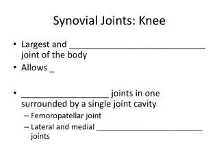
Types of Synovial Joints. Selected Synovial Joints: The Knee. This is considered the most complex joint in the human body. It is actually considered three joints working together. The Knee. These are:

E N D
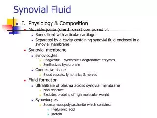
Selected Synovial Joints:The Knee This is considered the most complex joint in the human body. It is actually considered three joints working together.
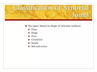
The Knee These are: • An intermediate joint between the patella and distal end of the femur (femoropatellar joint). This is a plane joint.
The Knee These are: • An intermediate joint between the patella and distal end of the femur (femoropatellar joint). This is a plane joint. • A lateral and medial tibiofemoral joints between the femoral condyles and the menisci below.
Tendon of quadriceps femoris Femur Suprapatellar bursa Articular capsule Patella Posterior cruciate ligament Subcutaneous prepatellar bursa Synovial cavity Lateral meniscus Lateral meniscus Infrapatellar fat pad Anterior cruciate ligament Deep infrapatellar bursa Tibia Patellar ligament (a) Sagittal section through the right knee joint Figure 8.8a The knee joint.
The Knee The menisci help prevent lateral motion and attach to the outer margins of the joint capsule on the tibia. They are easily torn.
Anterior Anterior cruciate ligament Articular cartilage on lateral tibial condyle Articular cartilage on medial tibial condyle Lateral meniscus Medial meniscus Posterior cruciate ligament (b) Superior view of the right tibia in the knee joint, showing the menisci and cruciate ligaments Figure 8.8b The knee joint.
The knee is unique in that it is not completely enclosed by a capsule.
The knee is unique in that it is not completely enclosed by a capsule. The articular capsule is found only on the lateral and posterior surfaces.
The knee is unique in that it is not completely enclosed by a capsule. The articular capsule is found only on the lateral and posterior surfaces. The anterior surface is covered by three ligaments going from the patella to the tibia.
These ligaments are: • The patella ligament
These ligaments are: • The patella ligament & • The medial and lateral patellar retinacula ligaments. They merge with the articular capsule on each side.
The intracapsular ligaments are the cruciate ligaments. The anterior and posterior cruciate ligaments cross each other forming an X in the notch between the femoral condyles.
The intracapsular ligaments are the cruciate ligaments. The anterior and posterior cruciate ligaments cross each other forming an X in the notch between the femoral condyles. They prevent anterior and posterior displacement.
Two additional ligaments, the Fibular and Tibial Collateral Ligaments prevent lateral or medial rotation when the knee is extended.
Quadriceps femoris muscle Tendon of quadriceps femoris muscle Patella Medial patellar retinaculum Lateral patellar retinaculum Tibial collateral ligament Fibular collateral ligament Patellar ligament Tibia Fibula (c) Anterior view of right knee Figure 8.8c The knee joint.
Medial femoral condyle Anterior cruciate ligament Medial meniscus on medial tibial condyle Patella (f) Photograph of an opened knee joint; view similar to (e) Figure 8.8f The knee joint.
The synovial cavity of the knee has a complicated shape and over one dozen associated bursae. Some are easily injured such as the subcutaneous prepatellar bursa which lies just over the patella (house maid’s knee).
Knee Injuries Common knee injuries involve the 3 C’s: • Collateral ligaments,
Knee Injuries Common knee injuries involve the 3 C’s: • Collateral ligaments, • Cruciate ligaments and
Knee Injuries Common knee injuries involve the 3 C’s: • Collateral ligaments, • Cruciate ligaments and • Cartilage (menisci).
Knee Injuries Lateral blows are the most dangerous, tearing the tibial collateral ligament and the medial meniscus and the anterior cruciate ligament.
Lateral Medial Patella (outline) Hockey puck Tibial collateral ligament (torn) Medial meniscus (torn) Anterior cruciate ligament (torn) Figure 8.9 A common knee injury.
Arthroscopic Knee Surgery Arthroscopy is a common surgical procedure in which a joint (arthro-) is viewed (-scopy) using a small camera.
Arthroscopic Knee Surgery Arthroscopy gives doctors a clear view of the inside of the knee. This helps them diagnose and treat knee problems.
Knee Replacement People with degenerative arthritis, chronic injuries often lose that smooth articular cartilage. The result is bone on bone. The knee joints must be replaced.
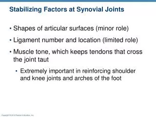
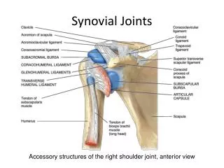
Shoulder (Glenohumeral) Joint The large head of the humerus fits into the glenoid cavity of the scapula. The cavity is extended by a fibrocartilage ring called the glenoid labrum. Connective tissue support comes from three groups of ligaments.
Acromion Coracoid process Coracoacromial ligament Articular capsule reinforced by glenohumeral ligaments Subacromial bursa Coracohumeral ligament Subscapular bursa Greater tubercle of humerus Tendon of the subscapularis muscle Transverse humeral ligament Scapula Tendon sheath Tendon of long head of biceps brachii muscle (c) Anterior view of right shoulder joint capsule Figure 8.10c The shoulder joint.
Shoulder (Glenohumeral) Joint B) Three Glenohumeral ligaments strengthen the front of the capsule. These ligaments are weak.
Acromion Coracoid process Articular capsule Glenoid cavity Glenoid labrum Tendon of long head of biceps brachii muscle Glenohumeral ligaments Tendon of the subscapularis muscle Scapula Posterior Anterior (d) Lateral view of socket of right shoulder joint,humerus removed Figure 8.10d The shoulder joint.
Shoulder (Glenohumeral) Joint C) The Rotator Cuff is formed from four tendons and muscles that encircle the joint. The muscles include the Subscapularis, Supraspinatus Infraspinatus and Teres minor.
Shoulder InjuriesRotator Cuff Because of its mobility, the stability of the shoulder joint has been sacrificed. Anterior dislocations are the most common along with damage to the rotator cuff muscles due to severe circumduction.
Shoulder InjuriesRotator Cuff Problems with the rotator cuff muscles can be classed into two categories – Tears of the tendons/muscles, and inflammation of the tendons (often called tendinopathy or tendonitis).
Elbow Joint This is a hinge joint where the radius and ulna articulate with the condyles of the humerus. The ulna’s trochlear notch forms a tight hinge with the trochlear of the humerus. This articulation allows for flexion and extension only.
Elbow Joint Side to side movement is prevented by the ulnar collateral ligament (triangular) and radial collateral ligament.
Articular capsule Anular ligament Humerus Coronoid process Medial epicondyle Ulnar collateral ligament Radius Ulna (d) Medial view of right elbow Figure 8.11d The elbow joint.
Humerus Anular ligament Medial epicondyle Radius Ulnar collateral ligament Articular capsule Coronoid process Ulna (c) Cadaver photo of medial view of right elbow Figure 8.11c The elbow joint.
Humerus Anular ligament Radius Lateral epicondyle Articular capsule Radial collateral ligament Olecranon process Ulna (b) Lateral view of right elbow joint Figure 8.11b The elbow joint.
Tommy John Surgery This procedure, more formally known as UCL (Ulnar Collateral Ligament) reconstruction, is designed to repair a torn elbow ligament—an injury typically caused by strong, repetitive overhead throwing motions of the arm.