HYDRONEPHROSIS
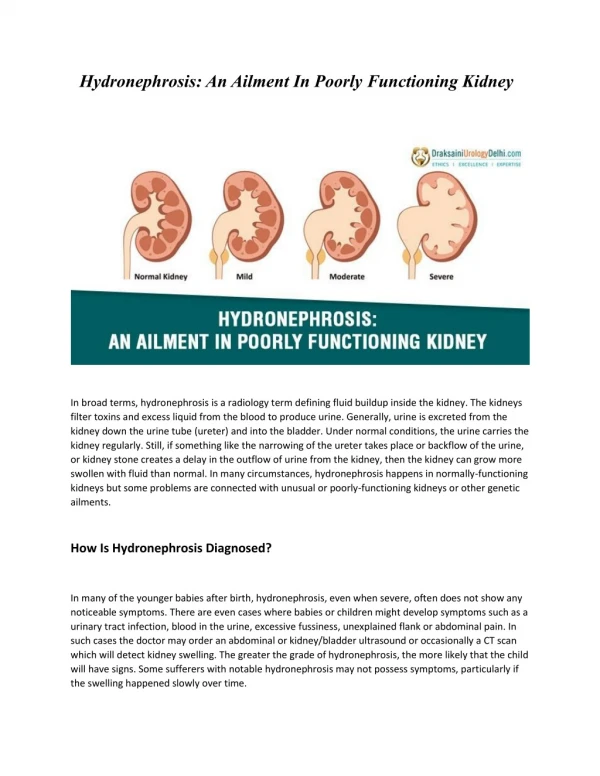
HYDRONEPHROSIS. Donna C. Queyquep, M.D. PGY II, Pediatrics November 19, 2004. Definition:. The dilation of the renal pelvis and calyces. May be considered a physiologic response to the interruption of urine. Not always caused by obstruction. Incidence:. Pre-natal ultrasound

HYDRONEPHROSIS
E N D
Presentation Transcript
HYDRONEPHROSIS Donna C. Queyquep, M.D. PGY II, Pediatrics November 19, 2004
Definition: • The dilation of the renal pelvis and calyces. • May be considered a physiologic response to the interruption of urine. • Not always caused by obstruction.
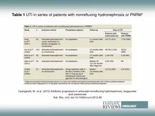
Incidence: • Pre-natal ultrasound • detects fetal anomaly in 1% of pregnancies, of which 20-30% are genitourinary in origin and 50% manifest as hydronephrosis
Causes: • Can be intrinsic, extrinsic or functional and can be classified as to level within the urinary tract • Ureter • Intrinsic • congenital ureteropelvic junction stricture • papillary necrosis • iatrogenic • blood clot • ureteral tumor
Causes: • Extrinsic • retroperitoneal cancer • aortic aneurysm • retrocaval ureter • inflammatory bowel disease • retroperitoneal hemorrhage • lymphocele
Causes: • Functional • gram-negative infection • neurogenic bladder • Bladder • Intrinsic • calculi • bladder neck contracture • Functional • VUR • neurogenic bladder
Causes: • Urethra • Intrinsic • urethral stricture • Extrinsic • BPH
Antenatal Period • The most common cause is physiologic dilation. • Metanephric urine production begins at 8 weeks, even before ureteral canalization is complete. • Transient obstruction with hydronephrosis occurs.
Pathophysiology: • Anatomic and functional processes interrupts the flow of urine. • There is a rise in ureteral pressure causing stretching and dilation; if pressures continue to rise, leads to decline in renal blood flow and GFR. • When significant obstruction is persistent, it affects nephrogenic tissue and results in varying degrees of cystic dysplasia and renal impairment.
Most Common Causes in Neonates: • Ureteropelvic Junction Obstruction • Ureterovesical Junction Obstruction • Posterior Urethral Valves • Eagle-Barrett Syndrome (a.k.a. Prune Belly Syndrome) • Vesicoureteral Reflux • Ureterocele
Ureteropelvic Junction Obstruction • UPJ is the most common cause of hydronephrosis in children. • May be the result of incomplete racanalizaton of the proximal ureter, abnormal development of ureteral musculature, abnormal peristalsis, ureteral valves or polyps. • Causes functional obstruction.
Ureteropelvic Junction Obstruction: • Male:female ratio is 2:1. • Prior to prenatal screening, about 25% were diagnosed in the first year of life. • Decreasing frequency with age.
Clinical Details of UPJ Obstruction: • Anatomically indistinct segment of the upper collecting system where the renal pelvis funnels into the ureter. • In 25-40% of kidneys, a supernumerary artery crosses the collecting system on its course into the kidney’s lower pole causing mechanical obstruction. • Occurs more often on the left side than on the right with a 3:2 ratio.
Other facts on UPJ: • Often associated with other congenital renal anomalies • ectopic or horseshoe kidneys • duplication of the collecting system • contralateral renal dysplasia • MCDK • renal agenesis • VUR (<40%), often low grade • VATER Syndrome
Diagnostic Modality for UPJ: • Ultrasonography is the initial modality of choice. • IVP • Retrograde pyelography • Radionuclide renogram
Assessments on UTS: • Renal length/size • Degree of caliectasis and parenchymal thickness • Presence of ureteral dilatation
Posterior Urethral Valves • Abnormal congenital mucosal folds that are thin membranes impeding bladder drainage. • Most common obstructive urethral lesion in male newborns found at the distal prostatic urethra. • Incidence is approx’ly 1 in 8,000 males. • Approx’ly 50% have reflux. • VCUG is the modality of choice.
Radiographic signs of PUV: • distended prostatic urethra • valve leaflets • bladder and/or bladder neck hypertrophy • diverticula • narrow stream in the penile urethra • incomplete emptying of the bladder
Treatment of PUV: • Transurethral valve ablation, vesicostomy or upper tract diversion • Urethral stricture is a common complication • Fetal intervention carries a high risk with mortality rate of 43% • ESRD, renal insufficiency and chronic renal failure are long-term consequences
30% of boys with posterior urethral valves whose symptoms present in infancy are at risk for progressive renal insufficiency.
Eagle-Barrett Syndrome • More commonly known as Prune Belly Syndrome • Characterized by: • deficiency of abdominal wall musculature • a dilated, non-obstructed urinary tract • bilateral cryptorchidism • talipes equinovarus and hip dislocation • Incidence is 1/35-50,000 • >95% occur in males
Believed to be caused by in-utero urinary tract obstruction and a specific mesodermal injury between the 4th and 10th week of gestation. Associated with renal dysplasia or agenesis. Often presents with a large-capacity, poorly contractile bladder. Heart, pulmonary, GI and orthopedic anomalies occur in a large percentage of PBS patients.
Management: • Neonatal Period • Optimize urinary tract drainage • Monitor and treat renal insufficiency • Antibiotic prophylaxis if reflux is present • Children • Surgical repair of reflux • Orchiopexy • Reconstruction of the abdominal wall • Renal transplant
Vesicoureteral Reflux • Retrograde propulsion of urine into the upper urinary tract during bladder contraction. • Primary reflux is caused by attenuation of the trigone and the contiguous intravesical ureteral musculature. • May be caused by the ectopic insertion of the ureter into the bladder wall resulting in a shorter intravesicular ureter, which acts as an incompetent valve during urination.
The ratio of the submucosal tunnel length to the ureteral diameter is the primary factor determining the effectiveness of the normal valve mechanism. • It is normally 5:1, and in those with reflux it is 1.4:1. • The intramural length increases from 0.5 cm at birth to 1.3 cm by 12 years of age. • Duplication of the collecting system and ureteroceles should also be considered.
Some clinical facts about VUR: • It is genetic. • Occurs in about 30% of first-degree relatives. • 1/3 of children with a urinary tract infection has reflux on VCUG. • Primary reflux tends to resolve over time as intravesical segment elongates with growth.
Resolves spontaneously before adolescence in: 90% of Gr. 1 reflux 80% of Gr. 2 50% of Gr. 3 10% of Gr. 4 0 in Grade 5 reflux Kidney is most susceptible to scarring in the first year of life and at the time of first upper tract infection. Scars less frequently develop after the age of 5. VUR and scarring lead to hypertension, progressive renal insufficiency and failure. Prognosis:
Treatment: • Observation • Medical treatment of infections • Surgical treatment • significant hydroureteronephrosis • indicated if impossible to keep urine sterile and reflux persists • acute pyelonephritis occurs • evidence of increasing renal damage
On Presentation, how do we manage? • Antenatal diagnosis of HN • Enlarged palpable kidneys on PE • Incidental finding on UTS done for other anomalies.
Management would depend on the clinical condition of the patient and the suspected nature of the lesion. • More common to have a unilateral HN that is not associated with systemic or pulmonary complications. • Postnatal UTS confirmation at about 1 month of life, depending on severity. • Bilateral HN • urgent work-up especially when accompanied by oligohydramnios and pulmonary disease • if male infant, postnatal VCUG and UTS
Prophylactic antibiotics (Amoxicillin 20mg/kg PO daily) before VCUG is performed, as hydronephrosis may be due to reflux. • DMSA scan to evaluate renal function. • Definitely: • Check presence and regularity of voiding. • Mild HN: UA, BMP • Moderate: UA, BMP, VCUG • Refer to specialist
References: • Tanagho and McAninch. Smith’s General Urology.16th ed: 2004, McGraw-Hill Companies, USA. • Zitelli and Davis. Atlas of Pediatric Physical Diagnosis. 4th ed:2002, Mosby, Inc., USA. • Resnick and Novick. Urology Secrets. 3rd ed:2003, Hanley and Belfus, Inc., USA. • Edmondson, J.D. Antenatal Hydronephrosis. Mar 2004, Emedicine article. • Wiener, J.S. Ureteropelvic Junction Obstruction, Congenital. Aug 2004, Emedicine article. • Maniam, P. Hydronephrosis and Hydroureter. Aug 2004, Emedicine article. • Div of Urology, Children’s Hosp, Boston, MA. Neonatal Hydronephrosis. Digital Urology Journal. Internet download.
References: • McCarthy, K. Vesicoureteral Reflux. Mar 2004, Emedicine article. • Behrman, et al. Nelson Textbook of Pediatrics. 17th ed: Saunders Co., USA. • Lloyd-Davies, et al. Color Atlas of Urology. 2nd ed:Mosby-Year Book Europe Limited, London, England. • Gillenwater, et al. Adult and Pediatric Urology. 4th ed, Vol. I: 2002, Lippincott Williams and Wilkins, USA. • Cloherty, et al. Manual of Neonatal Care. 5th ed: 2004, Lippincott Williams and Wilkins, USA. • Gonzales and Bauer. Pediatric Urology Practice. 1999: Lippincott Williams and Wilkins, USA.