DIAGNOSIS
DIAGNOSIS. Diagnosis of neurocysticercosis stems from suspicions that arises from the clinical manifestations of the disease. Most useful diagnostic test and the primary diagnostic criteria is neuroimaging 1. Contrast CT 2. MRI

DIAGNOSIS
E N D
Presentation Transcript
Diagnosis of neurocysticercosis stems from suspicions that arises from the clinical manifestations of the disease
Most useful diagnostic test and the primary diagnostic criteria is neuroimaging 1. Contrast CT 2. MRI • Useful in the evolution of cysticercus in th eparenchyma of brain.
4 Phases • Vesicular • Colloidal • Nodular-granular • Calcified phase
When only one cyst is seen in the transitional phase, it corresponds to the so-called "single enhancing lesion on CT" (SECTL), signifying a special syndrome.
Immunologic Assay • Enzyme ImmunoBlot • The current serological assay of choice for the diagnosis of neurocysticercosis • CDC's immunoblot is based on detection of antibody to one or more of 7 lentil-lectin purified structural glycoprotein antigens from the larval cysts of T. solium in an immunoblot format. • It is 100% specific. • No serum samples from patients with other microbial infections react with any of the T. solium-specific antigens. • Cumulative clinical experience has confirmed that in patients with multiple (more than two) lesions, the test has more than 95% sensitivity.
ELISA - Lack of specificity has been a major problem because of cross-reacting components in crude antigens derived from cysticerci - these components react with antibodies specific for other helminthic infections, especially echinococcosis and filariasis. - lower sensitivity than crude antigens and do not necessarily achieve higher specificity Assays employing crude antigens for the detection of antibody are not reliable for the identification of this disease
Diagnostic Criteria Absolute criteria • Histologic demonstration of parasite • Direct visualization of parasite by fundoscopic examination. • Evidence of cystic lesions showing scolex on CT/MRI. Major Criteria • Lesions suggestive of neurocysticercosis on neuroimaging studies • Positive immunological tests • Plain X-ray films showing calcifications in thigh and calf muscles Minor criteria • Subcutaneous nodules • Soft tissue or intracranial calcification on plain x-ray • Clinical manifestations suggestive of neurocysticercosis • Disappearance of intracranial lesions after a trial with anticysticercal drug
Epidemiologic • Living or coming from endemic area • Frequent travel to endemic areas • Household contact with Taeniasolium infection Definitive: 1 absolute 2 major 1 major + 2 minor + 1 epidemiologic Probable: 1 major + 2 minor 1 major + 1 minor + 1 epidemiologic 3 minor + 1 epidemiologic Possible: 1 major 2 minor 1 minor + 1 epidemiologic
Revised Absolute criteria • Histologic demonstration of parasite • Direct visualization of parasite by fundoscopic examination • Evidence of cystic lesions showing scolex on CT/MRI. Major Criteria • Lesions suggestive of neurocysticercosis on CT or MRI • Positive serum EITB (Enzyme Immunoblot Assay) • Resolution of cyst after therapy. • Spontaneous resolution of single enhancing lesions. Minor Criteria • Lesions compatible with neurocysticercosis on CT/MRI • Suggestive clinical features • Positive CSF ELISA • Cysticercosis outside CNS
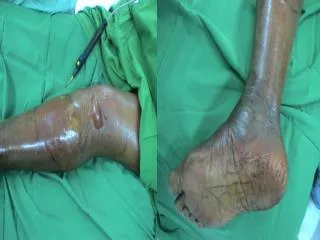
Definitive diagnosis of extra-neural cysticercosis will require one of the following: a) histopathological demonstration of parasite from excisional biopsy of a subcutaneous nodule. Demonstration of larval parts (hooks, suckers etc.) by fine needle aspiration cytology may provide a satisfactory alternative to open biopsy b) plain X-ray films showing multiple "cigar-shaped calcifications in the arm, thigh and calf muscles c) direct visualization of a cysticercosis larva in the anterior chamber of the eye with ultrasonography.
Epidemiologic • Living or coming from endemic area • Frequent travel to endemic areas • Household contact with Taenia solium infection Definitive: 1 absolute 2 major + 1 minor + 1 epidemiologic Probable: 1 major + 2 minor 1 major + 1 minor + 1 epidemiologic 3 minor + 1 epidemiologic