Download
1 / 19
320 likes | 2.22k Vues
Radiographic Positioning for Barium Enema. Presented by Aries Paul Zeta, RRT Property of Davao Doctors College. 10 – Miller’s Routine Sequence of Radiographs. AP – to include flexures Left lateral rectum AP – 15 – 25 degs. Cephalic(CR) to include rectum.

E N D
Radiographic Positioning for Barium Enema Presented by Aries Paul Zeta, RRT Property of Davao Doctors College
10 – Miller’s Routine Sequence of Radiographs • AP – to include flexures • Left lateral rectum • AP – 15 – 25 degs. Cephalic(CR) to include rectum. • 15 – 25 degs.RPO – to include Left colic • Right lateral – to include rectum
Cont… • Prone PA – to include flexures • Prone PA with 15 – 25 degs caudal angulation (Angle Prone)– to include rectum. • 15 – 25 degs LPO- to include the right colic flexure. • Supine – AP tightly collimated ileocecal region proj. taken in 2 – 3 degs obliquity. • Using horizontal central ray, upright proj. of both flexures and lateral rectum.
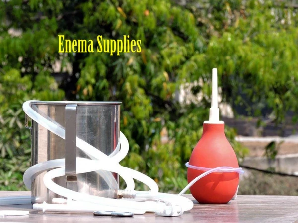
Modification of Positions for Barium Enema Usually used in the hospital
Scout Film First exposure of the procedure should be a plain radiograph of the abdomen area. Advice the patient to lie down on the radiographic table, the MSP of the patient should be inline with the MSP of the Table. Center the CR at the level of the L4 or the level of the iliac crest. Respiration is suspended during expiration. L4
Sim’s Position Sims position – relaxes the abdominal muscles and decreases pressure within the abdomen. Instruct the patient to lie on their side away from the tech. let the lower arm of the patient to be put at the back of his body. The up side knee should be flex for support and lower side extremity should not be flex or bent. Wearing gloves, coat enema tip with water-soluble lubricant.(KY jelly or any sterile lubricant). On expiration, direct enema tip toward the umbilicus proximally 1 to 1.5 inches. After initial insertion, advance up superiorly and slightly anteriorly. Do not force enema tip. Tape tubing in place to prevent slippage. Do not inflate unless directed by radiologist. Ensure IV pole/enema bag is no more than 24 inches (60cm) above the table. Ensure tubing stopcock is in the closed position and no barium flows into the pt.
Left/Right position of the recto sigmoid areaFilm: 10x12cm lengthwise True lateral position of the Recto sigmoid CR should be 5-7cm above the level of the pubic symphysis in the midaxillaryplane
AP (recto sigmoid area)Film: 10x12cm crosswise AP view of the Rectum & Sigmoid should be included CR 5-7 cm above the level of the pubic symphysis 5-7cm above pubic symphysis
AP (Single Contrast) Film: 14x17cm An Entire colon filled with contrast media should be demonstrated including the splenic flexure and the rectum. CR is at the level of the L4 or at the level of the iliac crest L4
AP Double ContrastFilm: 14x17cm lengthwise Patient lies in a supine position MSP is in line with the MSP of the table An Entire colon filled with positive and negative contrast media should be demonstrated including the splenic flexure and the rectum. CR is at the level of the L4 or at the level of the iliac crest L4
RPO Position(optional)Film: 14x17cm lengthwise Instruct the patient to lie on his right side making an angulation of 35-45deg It is taken primarily to demonstrate the Left Colic(splenic) flexure and ascending colon should be visualized. CR is at the level of the L4 or at the level of the iliac crest
LAO Position (optional)Film: 14x17cm lengthwise It is taken primarily to demonstrate the right colic (hepatic) flexure and sigmoid portion of the colon CR is at the level of the L4 or at the level of the iliac crest
Right Lateral DecubitusFilm: 14x17cm lengthwise Best demonstrate the “up” medial side of the ascending colon and the lateral side of the descending colon, when the colon is inflated with air due to gravity. CR at the level of the L4 or at the level of the iliac crest
Left Lateral DecubitusFilm: 14x17cm lengthwise Best demonstrate the “up”, medial side of the descending colon and the lateral side of the ascending colon, when the colon is inflated with air. CR is at the level of the L4 or at the level of the iliac crest
Ventral DecubitusFilm: 10x12cm lengthwise A cross table view of the recto sigmoid area Demonstrate the air-fluid level of the recto sigmoid area CR is at 5-7 cm above the level of the pubic symphysis in the midaxillary plane
PA Axial position (Angle Prone)Film: 10x12cm or 11x14cm crosswise Rectosigmoid area must be less superimposition than in the PA projection because of the angulation of the CR Center it the midline of the body with an angulation of 30-400caudad at approximate level of the anterior superior iliac spines.
Supine position Film: 14x17cm lengthwise A postevacuation radiograph view of the colon is taken after the procedure is done If inadequate satisfactory delineation of the mucus the patient may be given hot beverage (tea/coffee) to stimulate evacuation
After carePatient is advised to drink plenty of water, or laxative is taken to remove excess barium sulfate.
Acknowledgement • Radiographic positioning demonstrated by Davao Doctors College Interns