CELL MEDIATED IMMUNITY LABORATORY EVALUATION
420 likes | 582 Vues
CELL MEDIATED IMMUNITY LABORATORY EVALUATION. DEFICIENCIES: - PRIMARY (inherited) - SECONDARY (acquired) - infections (HIV) - malnutrition - trauma - cancer

CELL MEDIATED IMMUNITY LABORATORY EVALUATION
E N D
Presentation Transcript
CELL MEDIATED IMMUNITY LABORATORY EVALUATION
DEFICIENCIES: - PRIMARY(inherited) - SECONDARY(acquired) - infections (HIV) - malnutrition - trauma - cancer - therapy (radiation, cytostatics, steroids)
PRESENTATION: repeated severe infections of lung, skin, GIT, etc. caused by viral agents intracellular bacteria (Mycobacterium) opportunistic infections (Candida sp., Pneumocystis carinii)
DIAGNOSTIC PROCEDURES: are complex: patient’s and family history clinical evaluation laboratory tests: 1st level: leukocytes, differential cell count 2nd level: immunological testing
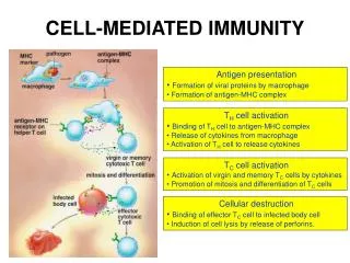
SKIN TEST IN VIVO: intradermal administration of anamnestic T-dependent antigens of microbial origin anamnestic Ag: previous contact is supposed natural exposition: candidin, toxoplasmin arteficial exposition: active immunisation (tetanus toxoid, PPD-purified proteine derivative of tuberculin, Mantoux reaction)
RESULTS: induration after 24 and 48 hours is read (DTH reaction) CD4+ T cells and macrophages infiltration (interferon ) INTERPRETATION: - positive: intact T cell immunity - negative: anergy
EX VIVO LABORATORY TESTS enumeration of various populations of immune cells functional tests
ENUMERATION OF IMMUNE CELLS cytomorphological criteria are not sufficient identification distinct cell population by the detection of specific surface molecules detection is based on specific immunochemical reactions between monoclonal antibodies and their targets (antigens) CD molecules: fully characterized leukocyte molecule
IMMUNOFLUORESCENCE ANALYSIS (IFA) antibody is labeled by fluorochromes Fluorochromes: - FITC : emission at 450 nm (green) - PE (phycoerythrin): emission at 580 nm (red) UV ABSORPTION FLUOROCHROME EMISSION laser (excitation) (light)
IMMUNOFLUORESCENCE ANALYSIS DIRECT IMMUNOFLUORESCENCE ANALYSIS: - primary antibody is labeled - whole blood - flow cytometry INDIRECT IMMUNOFLUORESCENCE ANALYSIS: - primary antibody is unlabeled - isolated cells, tissues - UV microscopy
DIRECT DOUBLE IMMUNOFLUORESCENCE TO DETECT CD3+ CD4+ T CELLS INDIRECT IMMUNOFLUORESCENCE TO DETECT CD3+ T CELLS Y Y ANTI CD3 FITC Mo Ab Y ANTI CD4 PE Mo Ab MOUSE ANTI CD3 Mo Ab INCUBATION RED CELLS LYSIS WASHING FLOW CYTOMETRY Y secondary Ab RaAMIgFITC INCUBATION INCUBATION RED CELLS LYSIS WASHING FITC WASHING Y Y FITC Y CD3 FITC Y Y Y Y TS/C CD3 CD3 FITC CD3 CD3 TS/C Y TH TH TH CD8 CD3 CD8 TS/C CD4 Y B B CD4 CD4 CD8 CD19 CD19 PE B CD19 FLOW CYTOMETRY
separation fluid (1,076 g/ml)
plasma LYMPHOCYTES separation fluid ERYTHROCYTES GRANULOCYTES
FLOW CYTOMETRY the most modern approach of cell analysis cells are directed in sheath flow of fluid CYTOMETRY DETERMINES: - number of cells - size of cells (FS parameter) - morphology of cells (SS) - the presence of fluorescence signal (FL) data are processed in PC MATERIAL EXAMINED: - whole blood, bone marrow - isolated cell suspensions - other body fluids (liquor, ascites, exudates) - cell suspension obtained by tissue desintegration CELLS ARE LABELED BY IMMUNOFLUORESCENCE (either direct or indirect) monoclonal antibodies are directed against - surface molecules - cytoplasmatic molecules - nuclear molecules
IMMUNOFLUORESCENCE ANALYSIS • OF THE WHOLE BLOOD • cell separation is ommited • direct immunofluorescence • heparinized blood is incubated with fluorochrome • labeled monoclonal antibody (FITC, PE) • hypotonic lysis of erythrocytes • removal of unbound monoclonal antibodies by washing • measurement by flow cytometry • Simultaneous determinantion of two (three) different molecules • is allowed by the application of two different monoclonals labeled • with different fluorochromes.
APPLICATION OF FLOW CYTOMETRY • IN CLINICAL MEDICINE • determination of different populations of leukocytes (other cell types) • immunophenotyping analysis of blood malignancies • determination of proliferating activity of cells: • - DNA content is estimated • - DNA is stained by intercalar chemicals which are fluorescent • determination of O2- dependent mechanisms of killing of phagocytes: • - granulocytes are incubated with chemical compound which is non • fluorescent • - oxygen species which are raised by NADPH oxidase change this • compound into fluorescent • cell ploidy determination • determination of intracellular biologically active molecules: • - cytokines (TH1, TH2 subsets) • - molecules regulating cell cycle • - molecules regulating cell apoptosis • - molecules encoded by oncogenes and antioncogenes
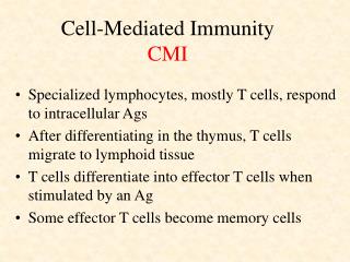
THE MOST IMPORTANT POPULATIONS OF PERIPHERAL BLOOD CD3+ T cells: population of mature T cells 50 - 75% CD4+ T cells: subpopulation of helper inducer T cells 30 - 60% CD8+ T cells: subpopulation of suppressor cytotoxic T cells 15 - 30% CD25+ T cells: activated mature T cells 1 - 5% CD19+ B cells: population of mature B cells 5 - 15% CD56+ NK cells: natural killers 5 - 15% CD14+ cells: monocytes CD15+ cells: granulocytes CD38+ cells: plasma cells
PRINCIPLES OF FLOW CYTOMETRY LABELED CELLS SENSORS DETECTING: NUMBER OF CELLS CELL SIZE (FS) CELL „ MORPHOLOGY“(SS) FL1 – GREEN EMISSION (FITC) FL2 – RED EMISSION (PE)
CELL SCATTERGRAM OF PERIPHERAL BLOOD granulocytes FS monocytes lymphocytes debris SS CD3-FITC CD3-FITC
DETERMINATION OF CD8+ T CELLS DETERMINATION OF CD4+ T CELLS CD8-PE CD4-PE CD3-FITC CD3-FITC
SARCOIDOSIS - BALF 55% lymphocytes
SARCOIDOSIS - BALF 92% CD3+ T-lymphocytes 8% CD8+ T-lymphocytes 85% CD4+ T-lymphocytes
1% lymphocytes PNEUMONIA - BALF 98% neutrophils
76% CD15 granulocytes 22% macrophages PNEUMONIA - BALF 4% CD19+ lymphocytes
FUNCTIONS OF LYMPHOCYTES IN VITRO • Functional capacity is tested as: • capacity of lymphocytes to proliferate • production of cytokines • Proliferation test • in vitro cultivation • cultivating media: supplemented by thymic factors • by antibiotics • buffer system • CO2 atmosphere
STIMULATION IS NECESSARY FOR LYMPHOCYTES PROLIFERATION ACTIVATORS: mitogens: - lectins from plants - polyclonal activators - specific binding to cell surface sugars - non-physiological proliferating stimulus - phytohemaglutinin (PHA) antigens: - specific stimulation of the single clone of T cell through TcR activators of cell kinases (phorbol esters)
CONCLUSION Cell proliferation is measured by the incorporation of isotope labeled nucleotides (3H thymidine) into newly formed DNA. Functional tests of T cell system are COMPLEMENTARY to the enumeration of cells.
LYMPHOCYTE PROLIFERATION IN VITRO lymphocytes in complete medium mitogen (phytohem- agglutinin) 3H ( )- thymidine 3H - thymidine uptake 96-wells panel well incubation 18h/37°C CO2 atmosphere incubation 72h/37°C CO2 atmosphere division DNA RADIOACTIVITY MEASUREMENT (cpm) CELL HARVEST (INCORPORATED RADIOACTIVITY)
DETERMINATION OF NATURAL CYTOTOXIC ACTIVITY NATURAL CYTOTOXIC CELLS: heterogenous population killing of viral infected and malignant cells the most important population are NK cells NK CELLS PHENOTYPE : CD56+ (NCAM-1)
NK CELL ACTIVITY: • the lytic activity of NK cells is tested • target cell line is labeled by radioactive Cr • target cells are mixed with isolated lymphoid cells • (NK cells are included) • during incubation target cells are lyzed • and released • Cr is measured • radioactivity release is the function • of NK cells activity
EVALUATION OF PHAGOCYTOSIS • absolute number of granulocytes (PMNL) • is limiting for succesful phagocytosis • four steps: priming, activation, adhesion • chemotaxis (oriented movement) • ingestion • killing, degradation
PRIMING, ACTIVATION, ADHESION OF GRANULOCYTES
IST STEP OF ADHESION, SELECTINS-MEDIATED collagens IMMUNOPATHOLOGY a1 b1 fibronectin a1 b1 INFECTION proinflammatory proadhesive signals a3 b1 MALIGNANCY MACROPHAGE CD62E IL-1 TNF CD34 CD62P E-CADHERIN CHEMOATTRACTANTS CD15 CD62L PECAM-1 PSGL-1 ENDOTHEL GRANULOCYTE rolling
2ND STEP OF ADHESION firm adhesion interaction between LFA-1 and ICAM-1 signaling: outside-in inside-out cell spreading 3RD STEP OF ADHESION diapedesis into tissues
4 b1 b2 II ND STEP OF ADHESION A C T I V A TION C-X-C CHEMOKINES ENDOTHELIA MACROPHAGES C-C CHEMOKINES ICAM-1 VCAM-1 ICAM-1 VLA-4 LFA-1 DIAPEDESIS EOSINOPHIL LFA-1 b2 NEUTROPHIL 3CYTOADHESINS FIBRINOGEN vWF
LABORATORY EVALUATION • presence of adhesion molecules is tested by immunofluorescence • LAD-II deficiency syndrome: • extremly rare • abnormal glycosylation of selectine ligands • LAD-I deficiency syndrome: • - absence of chain (CD18) of LFA-1 integrin heterodimers
CHEMOTAXIS • oriented movement of granulocytes in the gradient of chemoattractants • LABORATORY TEST: • migration of granulocytes under agarose layer • migration of granulocytes in Boyden’s chamber • DEFECTS OF CHEMOTAXIS: • primary defects: lazy leukocyte syndrome • - secondary defects: diabetes, juvenile periodontitis
INGESTION • „opsonins“ are necessary • incubation of heparinized blood with heat inactivated yeasts • granulocytes with ingested yeasts are read after staining under microscope lymphocyte erythrocyte yeast granulocyte with ingested yeasts
INTRACELLULAR KILLING • O2 - independent mechanisms: • immunochemical measurement of defensins • determinantion of lysosyme • INHERITED DEFECTS • deficiency of specific granules
INTRACELLULAR KILLING • O2 - dependent mechanisms: • INT (NBT) - test • isolated granulocytes are stimulated with starch granules in the presence of iodnitrotetrazolium salt (colorless) • generation of oxigen species (O2-., superoxide anion) by NADPH oxidases is the cause for the formation of formazan (red color) from INT • PRIMARY DEFECTS • CGD: - defects in NADPH oxidase assembly • - no formazan formation • SECONDARY DEFECTS • Associated with inflammatory response