Download
1 / 15
160 likes | 481 Vues
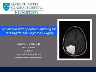
Intraoperative MRI Imaging Strategies to Evaluate for Complications during DBS Surgery. Olivia Huston Kendall Lee MD, PhD Robert Watson MD, PhD Matt Bernstein PhD John Huston MD Kiaran McGee PhD. Mayo Clinic Rochester, MN. Intraoperative MRI During DBS Surgery Purpose.

E N D
Intraoperative MRI Imaging Strategies to Evaluate for Complications during DBS Surgery Olivia Huston Kendall Lee MD, PhD Robert Watson MD, PhD Matt Bernstein PhD John Huston MD Kiaran McGee PhD Mayo Clinic Rochester, MN
Intraoperative MRI During DBS SurgeryPurpose To determine the optimal imaging sequences, the imaging findings and the clinical consequences of those findings for MRI performed during DBS surgery.
Intraoperative MRI During DBS SurgeryMethods • 143 patients underwent 152 DBS surgeries utilizing intraoperative 1.5T MRI. • MRI sequences utilized: - T1 MP-RAGE pre and intra-op - T2* GRE intra-op - T2 FLAIR selectively - T2 FSE selectively
Intraoperative MRI During DBS SurgeryResults Number of Surgeries: 152 • Subdural Hematomas: 5 • Subarachnoid Hemorrhages: 3 • Intraparenchymal Hemorrhage: 1 • Subarachnoid Air: 4 • Brain Shift: 144
Intraoperative MRI During DBS SurgerySubdural Hematoma • Number of SDH: 5 • Average thickness: 5.2 mm • Range: 4-8 mm
Intraoperative MRI During DBS SurgerySubarachnoid Hemorrhage • Number of SAH: 3 • One patient experienced headache and disorientation requiring 2 additional days of hospitalization. Symptoms cleared prior to discharge.
Intraoperative MRI During DBS SurgeryIntraparenchymal Hemorrhage • Number Hemorrhages: 1 • 5 x 5 mm
Intraoperative MRI During DBS SurgerySubarachnoid Air • Number of SA Air: 4
Intraoperative MRI During DBS SurgeryBrain Shift • Number with Shift: 144 • Average: 0.6 cm • Range: 0.1-1.3 cm
Intraoperative MRI During DBS SurgeryConclusions • Intracranial hemorrhage is occasionally identified but is rarely clinically significant. One delayed SDH required evacuation. • Brain shift during DBS surgery is common. • Subarachnoid air mimics subarachnoid blood on GRE. • Selective use of T2 FLAIR and T2 FSE imaging can confirm the presence hemorrhage or air and precludes the need for CT exams.