Download
1 / 23
230 likes | 465 Vues
SWIFT MRI for Breast Imaging. Curtis A. Corum , John C. Benson, Djaudat Idiyatullin, Angela Styczynski-Snyder, Diane Hutter, Lenore Everson, Lynn Eberly, Michael T. Nelson and Michael Garwood University of Minnesota Minneapolis, MN, USA RSNA 2012, VSBR51-04. What is SWIFT?.

E N D
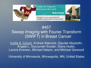
SWIFT MRI for Breast Imaging Curtis A. Corum, John C. Benson,Djaudat Idiyatullin, Angela Styczynski-Snyder, Diane Hutter, Lenore Everson, Lynn Eberly, Michael T. Nelson and Michael GarwoodUniversity of MinnesotaMinneapolis, MN, USA RSNA 2012, VSBR51-04
RSNA2012 VSBR51-04 Corum et al. SWIFT MRI for Breast Imaging What is SWIFT? SWeep Imaging with Fourier TransformDeveloped 2006 at University of Minnesota,Center for Magnetic Resonance Research
RSNA2012 VSBR51-04 Corum et al. SWIFT MRI for Breast Imaging What is SWIFT? ISMRM 2009 ISMRM 2010 JOE 2011
RSNA2012 VSBR51-04 Corum et al. SWIFT MRI for Breast Imaging Motivation Make more efficient use of scan time for Breast MRI by simultaneously obtaining: High Spatial Resolution Morphological Images and High Temporal Resolution DCE Images.
RSNA2012 VSBR51-04 Corum et al. SWIFT MRI for Breast Imaging Goals Optimize SWIFT sequence and protocol forBreast MRI at 4 T. Scan a pilot cohort of diagnostic patients. BI-RADS 4-5 Acquire simultaneously DCE and Morphological images. Optimize view sharing* based reconstruction. * Dougherty et al., (2007). High frame-rate simultaneous bilateral breast DCE-MRI., Magn Reson Med 57 : 220-225.
RSNA2012 VSBR51-04 Corum et al. SWIFT MRI for Breast Imaging SWIFT SWeep Imaging with Fourier TransformAlways Acquiring - Fast and Efficient short dead time - short T2 sensitive (2-6 μs) (preserve off-resonance) Smooth Gradient - Quiet and PNS free (peripheral nerve stimulation)
RSNA2012 VSBR51-04 Corum et al. SWIFT MRI for Breast Imaging SWIFT, Non-Cartesian... K-space Sampling Radial 3D, Center-Out 2 sec per 512 view sphere 3 sec with interleaved FS View Ordering (and gradient co-ordinate) One interleaved sphere (of many)
RSNA2012 VSBR51-04 Corum et al. SWIFT MRI for Breast Imaging SWIFT, Non-Cartesian....but... No gradient timing errors (group delay)... ...because gradient already ramped. Minimal eddy currents and thermal issues... ...because of small, smooth ramps. No special timing or k-space calibrations... ...either per scan or maintenance, or in post-processing.
RSNA2012 VSBR51-04 Corum et al. SWIFT MRI for Breast Imaging SWIFT Diagnostic Breast Protocol 2 min shimming, pre-scan, scout 20 sec SWIFT pre-scans, phase reference and gain 1-2 min SWIFT FOV check, FS (2-4 min) (optional) Double Angle Method GRE B1 map (2-4 min) (optional) SWIFT Variable Flip Angle T1 map 2-6 min SWIFT DCE FS, pre-contrast (MagnavistTM 0.1 mM/kg at 2 cc/s) 6 min SWIFT DCE FS post-contrast, (optional) further SWIFT test scans 11.33 min Minimum total timeOn Research Scanner, Agilent Console, Siemens Gradients, Oxford 4 T Magnet
RSNA2012 VSBR51-04 Corum et al. SWIFT MRI for Breast Imaging SWIFT Cases
RSNA2012 VSBR51-04 Corum et al. SWIFT MRI for Breast Imaging SWIFT Cases, subtraction MIPs 3D images on sized SWIFT compatible transmit/recieve single breast coils 256^3 matrix , isotropic < 1mm resolution OR 384^3 matrix, isotropic < 0.67 mm resolution (for more recent scans 10-14)
RSNA2012 VSBR51-04 Corum et al. SWIFT MRI for Breast Imaging Case 4, Mass Like DCIS 256^3 matrix , isotropic < 1mm (using all available data) Reformat to arbitrary slice without loss of image quality
RSNA2012 VSBR51-04 Corum et al. SWIFT MRI for Breast Imaging Case 4, Mass Like DCIS Clinical 1.5 T subtraction MIP High Resolution post contrast image is not at Maximum enhancement(DCE image is available) SWIFT 4 T Subtraction MIP High Resolution image at any time point is available
RSNA2012 VSBR51-04 Corum et al. SWIFT MRI for Breast Imaging Case 4, Mass Like DCIS MIP at each time point 6 sec frames MIP of all time points (maximum enhancement)
RSNA2012 VSBR51-04 Corum et al. SWIFT MRI for Breast Imaging Case 4, Mass Like DCIS DCE ROI 6 sec frames DCE ROI intensity 6 sec frames
RSNA2012 VSBR51-04 Corum et al. SWIFT MRI for Breast Imaging Case 12, FA 384^3 matrix , isotropic < 0.67mm (using all available data) Again, reformat to arbitrary slice without loss of image quality
RSNA2012 VSBR51-04 Corum et al. SWIFT MRI for Breast Imaging Case 13, IDC 384^3 matrix , isotropic < 0.67mm (using all available data) Again, reformat to arbitrary slice without loss of image quality
RSNA2012 VSBR51-04 Corum et al. SWIFT MRI for Breast Imaging Conclusion Demonstrated simultaneous high spatial resolution isotropic morphological and DCE Breast MRI with SWIFT.
RSNA2012 VSBR51-04 Corum et al. SWIFT MRI for Breast Imaging Breast SWIFT (near) Future... Dual Breast Coil Compressed Sensing Reconstruction Parallel Acceleration Siemens Research sequence implementation
RSNA2012 VSBR51-04 Corum et al. SWIFT MRI for Breast Imaging Acknowledgements We gratefully acknowledge NIH R21 CA139688, P41 RR008079, S10 RR023730, S10 RR027290,and the Minnesota Medical Foundation 3932-9227-09for grant support.Thanks to S. Suddarth and A. Rath of Agilent, B. Hannah,J. Strupp, and P. Anderson of CMRR for software and hardware support.Also thanks to Mike Tesch for assistance with the SWIFT distribution package, Jinjin Zhang for help with patients, and Carl Snyder and Gregor Adriany for assistance with coil modifications and design.
RSNA2012 VSBR51-04 Corum et al. SWIFT MRI for Breast Imaging SWIFT Parameters TR 4.4 ms, 62 kHz, 4.1 ms HS1, Flip 8-16 deg, 256 points Fat Suppression (FS) 1/8 views, 4 ms Gauss, Flip 120 deg, offset -625 Hz 3d Radial Isotropic Vieworder Sorted Halton** sequence, 512 views per k-space sphere 128 full spheres per 4.5 min acquisition (6 min with FS) 65,536 views total before restarting * 10 ms HS4 R20 pulse for dual fat and silicone suppression ** Wong TT, Sampling with Hammersley and Halton Points,J Graph Tools archive, Volume 2 , Issue 2, 1997. Chan RW et al., MRM 2010.
RSNA2012 VSBR51-04 Corum et al. SWIFT MRI for Breast Imaging NMR and Convolution x(t) RF pulse h(t) spin impulse response r(t) = system response * NMR and Convolution The fundamental basis of SWIFT signal processing is that a frequency modulated pulse alters the system response away from the familiar hard pulse impulse response. In the small flip angle limit the relationship is convolution. Practically it works well up to 90°.
RSNA2012 VSBR51-04 Corum et al. SWIFT MRI for Breast Imaging SWIFT and Correlation x(t) RF pulse r(t) system response h(t) spin impulse response = Recovering a standard FID by correlation SWIFT produces an FID if the raw data (system reposnse) is correlatied with the complex RF pulse shape as a post processing step. In practice this is performed in the frequency domain by multiplication with the complex conjugate of the complex pulse profile.