Download
1 / 32
320 likes | 340 Vues
Dive into the structure and functions of the female reproductive system, from ovary details to the menstrual cycle phases. Learn about oogenesis, uterine tubes, uterus, cervix, vaginal health, and external genitalia.

E N D
Martini’s Visual Anatomy and Physiology First Edition Martini w Ober Chapter 25Reproductive System – Female I Lecture 22
Lecture Overview • Functions of the female reproductive system • The ovaries – structure and function • Female internal reproductive organs • Female external reproductive organs
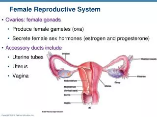
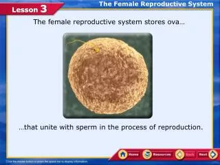
Functions of the Female Reproductive System • Produce and maintain sex cells (eggs) – a function of the ovaries, the primary sex organ • Transport eggs to site of fertilization • Produce female sex hormones • Provide favorable environment for development of offspring • Move offspring to outside (birth)
Organs of the Female Reproductive System (In anteflexion) (Skene’s glands; lesser vestibular glands) (Bartholin’s glands) Figure from: Martini, Anatomy & Physiology, Prentice Hall, 2001
Female Pelvic Cavity Figure from: Hole’s Human A&P, 12th edition, 2010
Ovary Attachments Figure from: Hole’s Human A&P, 12th edition, 2010 (Retracted) (Mesentery)
Ovaries and Their Attachments Oophorectomy – removal of one or both ovaries Fold of peritoneum that attaches to sides and floor of pelvic cavity (limits side-to-side movement and rotation) Posterior view Figure from: Martini, Anatomy & Physiology, Prentice Hall, 2001
Overview of Female Reproductive Cycle Figure from: Hole’s Human A&P, 12th edition, 2010
Overview of the Ovarian Cycle Ovarian cycle – events occurring monthly in an ovary (oocyte growth and meiosis occur); cycle is usually about 28 days long Figure from: Hole’s Human A&P, 12th edition, 2010 Two phases: 1) Follicular phase 2) Luteal phase
Oogenesis Oogonia = stem cells Process stops in meiosis I (Prophase) Stimulated by FSH/LH About 2 million primary oocytes at birth. By puberty, there are about 400,000. Fewer than 400-500 will be released during a female’s reproductive life. Probably fewer than 10 will be fertilized. How does oogenesis differ from spermatogenesis? How is it the same?
Ovarian Cycle – Preovulatory (Follicular) Phase Figure from: Martini, Anatomy & Physiology, Prentice Hall, 2001 (Graafian) 1.5 cm Many Few One Thecal and granulosa cells produce estrogens 8-10 days after beginning of cycle 10-14 days (FSH) (FSH) LH Meiosis II started Meiosis I Estrogen
Ovarian Cycle – Postovulatory (Luteal) Phase (Day 14) 12 days post ovulation Lipids used to synthesize progestins, e.g., progesterone (prepares uterine lining for implantation) LH If fertilization has not occurred LH Figure from: Martini, Anatomy & Physiology, Prentice Hall, 2001
Ovulation Figure from: Hole’s Human A&P, 12th edition, 2010
Female Internal Accessory Organs Figure from: Hole’s Human A&P, 12th edition, 2010
Uterine (Fallopian) Tubes Takes about 4 days for an oocyte to travel from the infundibulum to the uterine cavity Segments of the uterine tube: - Infundibulum contains fimbriae (inner surfaces lined with cilia that beat toward center) - Ampulla (middle, muscular segment) - Isthmus (segment connected to the uterine wall) Fertilization usually occurs around here Oocytes are transported by - ciliary action - peristalsis (Parasympathetic NS activity a few hours before ovulation) Fallopian tube = salpinx [salping(o)-] Figure from: Martini, Anatomy & Physiology, Prentice Hall, 2001
Lining of Uterine Tubes Figure from: Hole’s Human A&P, 12th edition, 2010 Tall ciliated columnar epithelial cells with interspersed mucin-secreting cells. Tubes contain glycoproteins and lipids
Uterus (hyster(o)-) - Mechanical protection (fetus) - Nutritional support (fetus) - Waste removal (fetus) - Ejection of fetus at birth Cervical mucous prevents spread of bacteria from vagina to uterus Figure from: Martini, Anatomy & Physiology, Prentice Hall, 2001
Uterine Wall Smooth muscle of myometrium is arranged in longitudinal, circular, and oblique layers Under the influence of estrogen, uterine glands, blood vessels, and epithelium in the functional zone of the endometrium change with the phases of the uterine (menstrual) cycle Figure from: Martini, Anatomy & Physiology, Prentice Hall, 2001
Clinical Application Cervical Cancer and Pap Smears (Cytology) Figures from: Saladin, Anatomy & Physiology, McGraw Hill, 2007 Cervical cancer is more common in: - Women between the ages of 30 and 50 - Women who smoke - Sexual activity at an early age/history of STDs or cervical inflammation (HPV)
Vagina Acidity of vagina protects adults from bacterial infections Major functions: - Passageway for elimination of menstrual fluids - Receives penis and holds sperm prior to passage into uterus - Inferior portion of birth canal for fetal delivery Figure from: Martini, Anatomy & Physiology, Prentice Hall, 2001
Female External Reproductive Organs Female external genitalia = pudendum or vulva anterior Includes the structures external to the vagina: - mons pubis - labia majora and minora - clitoris - vestibular structures Opening of ducts of greater vestibular glands (Bartholin’s) – mucous secretions Perineum Know the terms on this slide posterior Figure from: Martini, Anatomy & Physiology, Prentice Hall, 2001
The Deep Female Perineum Figure from: Saladin, Anatomy & Physiology, McGraw Hill, 2007
Development of External Reproductive Organs Figure from: Hole’s Human A&P, 12th edition, 2010
Erection, Lubrication, and Orgasm Figure from: Hole’s Human A&P, 12th edition, 2010
Review • Function of the female reproductive system • Produce and maintain sex cells (eggs) – a function of the ovaries, the primary sex organ • Transport eggs to site of fertilization • Provide favorable environment for development of offspring • Move offspring to outside (birth) • Produce female sex hormones
Review • Several ligaments hold female reproductive structures in place • Broad ligament • Suspensory ligament • Ovarian ligament • Uterosacral ligament • Peritoneum-lined recesses in female • Rectouterine pouch – separates uterus from colon • Vesciouterine pouch – separates uterus from urinary bladder
Review • During oogenesis • Oogonia stop development in meiosis I (before birth) • Secondary oocytes, rather than mature gametes, are released monthly at ovulation • Ovarian cycle • Cycle is about 28 days long • Two main phases • Preovulatory (follicular) – 14 days • Postovulatory (luteal) – 14 days
Review • Ovarian cycle (continued) • Preovulatory (follicular) phase • FSH stimulates primordial follicle to develop • Primary follicle secretes estrogen (granulosa and thecal cells) • Tertiary follicle is a mature (Graafian) follicle • Postovulatory (luteal) • LH stimulates rupture of tertiary follicle (ovulation) • Corpus luteum develops from remnants of follicle (granulosa cells) • Corpus luteum secretes progesterone which prepares the uterus for implantation • If pregnancy does not occur, corpus luteum involutes to become the corpus albicans (scar tissue)
Review • Female internal accessory organs • Uterus • Anteflexed muscular organ that will hold developing fetus • Body • Fundus is furthest away from vagina • Perimetrium • Myometrium (thick smooth muscle layer) • Endometrium (Functional zone, basilar zone) • Uterine (fallopian) tubes • Infundibulum (contains fimbriae) • Ampulla (thick muscular wall) • Isthmus (connection with uterus) • Fertilization usually occurs in the ampulla/isthmus boundary • Lined with cilia; smooth muscle to capture released oocyte • Nutrient-rich environment (lipids and glycogen)
Review • Female internal accessory organs (continued) • Vagina • Elastic, muscular tube between cervix and vestibule • Passageway for elimination of menstrual fluids • Receives penis and holds sperm prior to passage into uterus • Inferior portion of birth canal for fetal delivery • Maintains an acidic environment (in adults) to prevent infections • Parasympathetic stimulation – expansion and elongation during sexual stimulation • Vestibular glands along sides of vagina secrete mucus to lubricate the vaginal lumen
Review • Female External Genitalia • Entire area is the vulva or pudendum • Mons pubis, labia majora • Labia minora, vestibule • Anterior to posterior: clitoris, urethra, vaginal entrance • Bartholin’s glands (greater vestibular); ducts open just posterior to vaginal entrance • Skene’s glands (paraurethral, lesser vestibular); ducts open posterior to urethral meatus
Review • Perineum • Diamond-shaped area of the trunk between the thighs and buttocks extending from the pubis to the coccyx (between ischial tuberosities) • Shallow compartment lying between this diamond shaped area and the pelvic floor (formed by pelvic diaphragm) • Male perineum contains: penis, scrotum, anus • Female perineum contains: vulva, anus