FEMALE REPRODUCTIVE SYSTEM
FEMALE REPRODUCTIVE SYSTEM. FEMALE REPRODUCTIVE SYSTEM. TO REVIEW THE COMPONENTS OF THE FEMALE REPRODUCTIVE SYSTEM . TO CHARACTERIZE THE GENERAL ORGANIZATION OF THE OVARIES . TO UNDERSTAND THE HORMONAL REGULATION OF OOGENESIS, OVULATION, AND THE UTERINE CYCLE.

FEMALE REPRODUCTIVE SYSTEM
E N D
Presentation Transcript
FEMALE REPRODUCTIVE SYSTEM TO REVIEW THE COMPONENTS OF THE FEMALE REPRODUCTIVE SYSTEM TO CHARACTERIZE THE GENERAL ORGANIZATION OF THE OVARIES TO UNDERSTAND THE HORMONAL REGULATION OF OOGENESIS, OVULATION, AND THE UTERINE CYCLE
Hormones of the Female Reproductive Cycle • Control the reproductive cycle • Coordinate the ovarian and uterine cycles
Hormones of the Female Reproductive Cycle • Key hormones include: • FSH • Stimulates follicular development • LH • Maintains structure and secretory function of corpus luteum • Estrogens • Have multiple functions • Progesterones • Stimulate endometrial growth and secretion
FEMALE REPRODUCTIVE SYSTEM OVARIES OVIDUCT (UTERINE TUBES) UTERUS VAGINA
Female Reproductive Organs • Ovary: female gonad • Uterine Tubes (fallopian tube, oviduct) - three parts: infundibulum, ampulla, isthmus
The Female Reproductive System in Midsagital View Figure 28.13
The Ovaries and Their Relationships to the Uterine Tube and Uterus Figure 28.14a, b
The Uterine Tubes Figure 28.17a-c
The Uterus • Muscular organ • Mechanical protection • Nutritional support • Waste removal for the developing embryo and fetus • Supported by the broad ligament and 3 pairs of suspensory ligaments
Uterine Wall Consists of 3 Layers: • Myometrium – outer muscular layer • Endometrium – a thin, inner, glandular mucosa • Perimetrium – an incomplete serosa continuous with the peritoneum • The site of implantation of developing embryo • And 3 parts: fundus, body, and cervix
Female Accessory Sex Organs: Uterus • Uterine endometrium has two layers: - basal layer - functional layer: built up and shed each cycle
The Uterus Figure 28.18a, b
The Uterus Figure 28.18c
The Uterine Wall Figure 28.19a
The Uterine Wall Figure 28.19b
The Uterine CycleTo be discussed below Figure 28.20
Functions of the Ovary • Production of a mature oocyte, capable of fertilization and embryonic development. • Production of ovarian steroids (estradiol, progesterone). • Production of gonadal peptides (inhibin, activin).
Structural Organization of the Ovary • The main functional unit of the ovary is the follicle. • Follicles are composed of the oocyte, granulosa cells, and theca cells.
Stages of Follicular Growth • Follicles are present in a number of different stages of growth: - primordial follicles (resting) - primary, secondary, and antral follicles - preovulatory (Graafian) follicles
The Corpus Luteum • After the preovulatory follicle ovulates (releases its egg), it forms the corpus luteum.
FEMALE REPRODUCTIVE SYSTEM OVIDUCT (UTERINE TUBES) INFUNDIBULUM, AMPULLA, ISTHMUS, UTERINE UTERUS FUNDUS, BODY (CORPUS), CERVIX
Oogenesis and Oocyte Maturation • Recall that germ cells must go through meiosis in order to produce unique haploid cells. • From one spermatogonia, end up with four spermatozoa. • Oocytes must also go through meiosis, but they do it during the course of follicular development. • Primordial follicles contain primary oocytes that are arrested in prophase I, prior to the first meiotic division (diploid). • How do they go through the rest of meiosis?
LH surge zona pellucida first polar body Oocyte Maturation • Oocytes remain in prophase I until the preovulatory surge of LH, which initiates completion of the first meiotic division. • The primary oocyte does not split into two cells, but instead gives off a very small first polar body, containing half of the chromosomes.
fertilization first polar body first polar body second polar body Oocyte Maturation • Thus, the ovulated “egg” is actually not completely mature (hasn’t completed meiosis II). • Maturation goes to completion only if the oocyte is fertilized. • Fertilization causes completion of meiosis II, and expulsion of a second polar body. • Meiosis of the oocyte results in only one gamete.
FEMALE REPRODUCTIVE SYSTEM The Ovarian Cycle OVARY GERMINAL EPITHELIUM TUNICA ALBUGINEA - thin connective tissue capsule underlying germinal epithelium CORTEX - surrounds the medulla and contains maturing follicles MEDULLA - central connective tissue containing vascular supply and nervous innervation
FEMALE REPRODUCTIVE SYSTEM The Ovarian Cycle OVARY 3 to 5 million OOGONIA differentiate into PRIMARY OOCYTES during early development OOCYTES becomes surrounded by squamous (follicular) cells to become PRIMORDIAL FOLLICLES most PRIMORDIAL FOLLICLES undergo atresia leaving 400,000 at birth oocytes at birth arrested at Meiosis I (prophase)
OOGENESIS FEMALE REPRODUCTIVE SYSTEM OVARY THREE STAGES OF OVARIAN FOLLICLES CAN BE IDENTIFIED FOLLOWING PUBERTY: (each follicle contains one oocyte) (1) PRIMORDIAL FOLLICLES - very prevalent; located in the periphery of the cortex - a single layer of squamous follicular cells surround the oocyte (2) GROWING FOLLICLES - three recognizable stages: (a) early primary follicle (b) late primary follicle (c) secondary (antral) follicle (3) MATURE (GRAAFIAN) FOLLICLES - follicle reaches maximum size
FEMALE REPRODUCTIVE SYSTEM OVARIAN FOLLICLES (1) PRIMORDIAL FOLLICLES (2) GROWING FOLLICLES (a) early primary follicle - follicular cells still unilaminar but now are cuboidal in appearance - oocyte begins to enlarge (b) late primary follicle - multilaminar follicular layer; cells now termed granulosa cells - zona pellucida appears; gel-like substance rich in GAGs - surrounding stromal cells differentiate into theca interna and theca externa (b) secondary (antral) follicle - cavities appear between granulosa cells forming an antrum - follicle continues to grow - formation of cumulus oophorus and corona radiata (3) MATURE (GRAAFIAN) FOLLICLES
FEMALE REPRODUCTIVE SYSTEM OVARIAN FOLLICLES late primary follicle
FEMALE REPRODUCTIVE SYSTEM OVARIAN FOLLICLES GRANULOSA (FOLLICULAR) CELLS OOCYTE ZONA PELLUCIDA
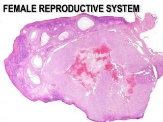
FEMALE REPRODUCTIVE SYSTEM OVARY CORTEX MEDULLA CORPUS LUTEUM
FEMALE REPRODUCTIVE SYSTEM OVARY CORTEX TUNICA ALBUGINEA GERMINAL EPITHELIUM PRIMORDIAL FOLLICLES
FEMALE REPRODUCTIVE SYSTEM OVARY TUNICA ALBUGINEA GERMINAL EPITHELIUM
FEMALE REPRODUCTIVE SYSTEM OVARY OVARY H&E PRIMORDIAL FOLLICLES EARLY 1º
FEMALE REPRODUCTIVE SYSTEM OVARY OVARY OVARY H&E CORPUS ALBICANS EARLY PRIMARY FOLLICLES PRIMORDIAL FOLLICLE
FEMALE REPRODUCTIVE SYSTEM OVARY LATE PRIMARY FOLLICLE multilaminar
FEMALE REPRODUCTIVE SYSTEM OVARY MATURE (GRAAFIAN) FOLLICLE zona pellucida cumulus oophorus corona radiata theca interna and externa theca interna cells begin to produce androgens that are converted to estrogens
FEMALE REPRODUCTIVE SYSTEM HORMONAL REGULATION OF OOGENSIS AND OVULATION HYPOTHALAMUS release of GnRF which stimulates release of LH and FSH from the adenohypophysis (ANTERIOR PITUITARY)
CNS hypothalamus GnRH Pituitary FSH LH OVARY Neuroendocrine Regulation of Ovarian Functions Follicle Development E2, P Ovulation inhibin, activin Luteinization
Effects of GnRH on Gonadotropins • GnRH is released in a pulsatile manner, stimulating the synthesis and release of LH and FSH. • GnRH acts through its receptor on the pituitary gonadotroph cells, stimulating production of phospholipase C. • Recall that IP3 pathway causes gonadotropin release, while the DAG/PKC pathway causes gonadotropin synthesis.
Actions of FSH on Granulosa Cells FSH AC ATP Gs cAMP PKA CREB CRE Gene Expression Steroidogenic enzymes LH Receptor Inhibin Subunits Plasminogen activator
Regulation of Estradiol Production • Recall the two-cell theory of estradiol production (lecture 4): - LH acts on theca cells to produce androgens - FSH acts on granulosa cells to increase aromatase activity, resulting in conversion of androgens to estrogens (granulosa cells lack 17 hydroxylase activity)
LH androgens Theca cells aromatase Granulosa cells FSH Ovarian Estradiol Production estradiol
Regulation of Progesterone Production • Progesterone is produced from theca cells, mature granulosa cells, and from the corpus luteum. • In this case, gonadotropins induce expression of - steroidogenic acute regulatory protein - P450 side chain cleavage
Actions of Estradiol • Estradiol plays an important role in feedback regulation of gonadotropin release. • Low estradiol levels exert negative feedback (via inhibition of GnRH release) Question: what happens to LH and FSH levels if you remove the ovary (increase, decrease, no change)? Answer:
Actions of Estradiol • High estradiol levels exert positive feedback (via increase in GnRH receptors, stimulation of GnRH release, increased pituitary response to GnRH, and effects on LHb) - increase in stimulatory neurotransmitters regulating GnRH neurons (ie, norepinephrine) - decrease in inhibitory neurotransmitters (ie, beta endorphin) - increased activity of GnRH neurons - increased expression of GnRH receptors - increased expression of LHb gene
Actions of Estradiol • Estradiol also has important actions in a number of other tissues: - causes proliferation of uterine endometrium - increases contractility of uterine myometrium - stimulates development of mammary glands - stimulates follicle growth (granulosa cell proliferation) - effects on bone metabolism, hepatic lipoprotein production, genitourinary tract, mood, and cognition • Effects are mediated through the intracellular estrogen receptors (alpha and beta), and possible membrane effects.
Actions of Progesterone • Progesterone exerts positive and negative feedback effects on gonadotropin synthesis and release. • Progesterone also acts on many tissues: - stimulates secretory activity of the uterine endometrium - inhibits contractility of the uterine myometrium - stimulates mammary growth • The actions of progesterone are mediated through an intracellular P receptor, which acts as a transcription factor.