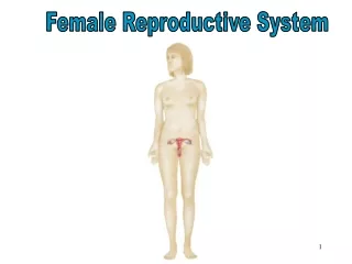
Female Reproductive System
This overview of the female reproductive system highlights the key components, including the ovaries, fallopian tubes, uterus, vagina, and external genitalia. The ovaries, almond-shaped structures, play a vital role in hormone production and the maturation of follicles. The uterus serves as the site for fetal development, with its layered structure designed for growth and menstruation. We explore the functions of the vagina, hormonal regulation of the menstrual cycle, and the impact of external factors on reproductive health.

Female Reproductive System
E N D
Presentation Transcript
Ovaries • 2 structures (L & R) • About 1.5 inches long • Shape is similar to almonds but larger • Produce what hormones? • Primary follicles – at birth (around how many?) • Only 300 – 400 become mature
Follicles • Graafian follicle – mature follicle • Atretic follicles – immature follicles that begin to deteriorate • Corpus luteum – ruptured follicle (released ovum for ovulation) & secretes progesterone & estrogen
Fallopian Tubes • About 4 inches long • Lateral end – ovaries • Medial end – uterus • Fimbriae– fringe – like projections that create currents in fluid to draw ovum into the tubes • Fertilization happens here (sperm & ovum meet) • What smooth muscle contraction moves the ovum through the tube?
Uterus • Shaped like an upside – down pear • Dimensions: 3 in x 2 in x 1 in • Superior to the bladder in which cavity? • Very muscular to stretch for a growing fetus • Layers: • Myometrium – smooth muscle of the outermost layer • Endometrium – inside layer; lining of the uterus • Functional layer – regenerated & lost during menstrual cycle
Anatomy of the Uterus • Fundus – upper portion above the fallopian tubes • (measured in pregnancy) • Body – large central part • Cervix – the opening from the vagina to body
Vagina • Muscular tube • 4 inches long • Posterior to the urethra & anterior to rectum • Hymen – covers the opening of the vagina • 3 functions: • Receive sperm from sexual intercourse • Provide exit for menstrual cycle • Provides a birth canal
Vagina cont. • Vaginal mucosa protects the female reproductive system from infection • Higher pH decreases bacterial growth • Normal flora – increases pH & prevent bacteria
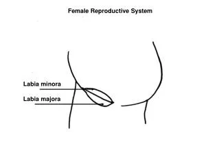
External genitalia • Vulva • Labia Majora • Labia Minora • Clitoris • Bartholin’s glands
Mammary Glands • Produce mild for offspring • Anterior to pectoralis major muscles • Alveolar glands – produce milk • Areola – dark pigmented area around nipple • What hormone controls milk production? • Which hormone controls milk release (“let down”)?
Menstrual cycle • Hormones • FSH - • Leutinizing Hormone • Estrogen • Progesterone
3 phases • Menstrual phase – menses or menstruation occurs • Lasts 2 – 8 days • FSH increases; estrogen & progesterone decreases • Follicular phase – Ovarian follicles mature • LH slowly increases • Estrogen increases to increase the endometrium to regenerate the functional layer • Luteal phase – rupture of follicle to release ovum • Corpus luteum forms • Progesterone increases functional layer
Other hormones • Inhibin – decreases FSH production • Relaxin – inhibits contractions of the myometrium to increases chances of implantation
Causes of irregular menstrual cycles • Amenorrhea • Decreases fat • Vigorous exercise • Extreme emotional states • Anorexia • Endocrine disorders