ENTEROPATHOGENIC ESCHERICHIA COLI II
750 likes | 1.09k Vues
ENTEROPATHOGENIC ESCHERICHIA COLI II. Hin-chung Wong Department of Microbiology Soochow University. SHIGA-LIKE TOXINS.

ENTEROPATHOGENIC ESCHERICHIA COLI II
E N D
Presentation Transcript
ENTEROPATHOGENICESCHERICHIA COLIII Hin-chung Wong Department of Microbiology Soochow University
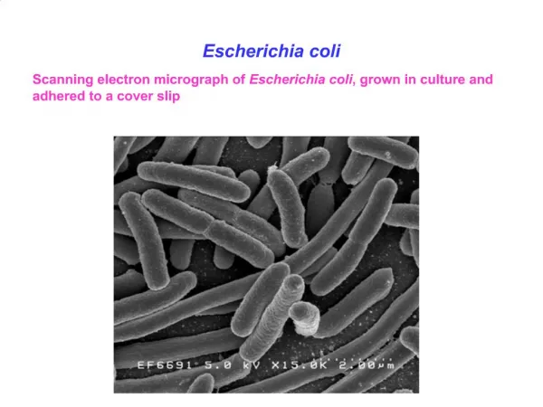
SHIGA-LIKE TOXINS • Enteropathogenic E. coli (EPEC, e.g. O26:H11, O128:B12, O138:K81) and enterohemorrhagic E. coli (EHEC, e.g. O157:H7) are not enteroinvasive and do not produce the classical heat-stable and heat-labile enterotoxins. • A cytotoxin known as Shiga-like toxin was demonstrated in EPEC and EHEC. • A cytotoxin which could be neutralized by anti-shiga toxin is known as Shiga-like toxin I (SLT-I), and the non-neutralizable one is known as shiga-like toxin II (SLT-II)
SHIGA-LIKE TOXINS • In Thailand, SLT-producing E. coli was isolated from 9% of market beef specimens, from 8 to 28% of fresh beef specimens at slaughterhouses, and from 11 to 84% of fecal specimens from cattle • Vero cell cytotoxin E. coli O157:H7 was isolated from 3.7% of beef, 1.5% of port, 1.5% of poultry, and 2% of lamb samples in Canada
SHIGA-LIKE TOXINS • The SLT-I is also known as verotoxin 1 and the SLT-II is also known as verotoxin 2. • E. coli producing large among of SLTs are also named as VTEC (Verotoxin producing E. coli)
SHIGA-LIKE TOXINS • Iron is known to depress Shiga toxin production by Shigella dysenteriae 1, and temperature has been shown to regulate several genes required for Shigella invasiveness and also expression of virulence plasmid in Yersinia. • Iron also suppressed SLT-I synthesis in E. coli lysogenized with phage 933J but did not demonstrably repress toxin synthesis in E. coli strains carrying the cloned slt-I genes (Table 3).
SHIGA-LIKE TOXINS • The SLT-I and SLT-II have been purified to homogeneity. • The bacteria grown in iron-depleted medium were disrupted by French press and the toxins purifed by anti-shiga toxin affinity chromatography or by conventional biochemical methods. • The SLT-I A subunit has a MW 32,200 and the SLT-I B subunit has a MW of 7,700
SHIGA-LIKE TOXINS • The SLT-II was purified from E. coli strain containing the cloned toxin genes on recombinant plasmid. • Purification was accomplished by a series of column chromatography techniques including monoclonal antibody affinity chromatography (against SLT-II). • The SLT-II consisted of A and B subunits with apparent molecular weights of 32,000 and 10,200, respectively.
SHIGA-LIKE TOXINS • By analogy with Shiga toxin, the most likely A-to-B subunit ratio for SLT-I is 1:5. • The MW of holotoxin is about 70,000
SHIGA-LIKE TOXINS • The purified SLTs have the same biological activities as and comparable specific activities to purified Shiga toxin. • The molecular basis is probably the catalytic inactivation of 60S ribosomes in toxin-sensitive (receptor-expressing) cells (Fig. 14) • The receptor for SLT-I and SLT-II has been identified and is the same as for Shiga toxin; it is a globotriosyl ceramide containing a galactose-à-(1->4)-galactose-á-(1->4)-glucose ceramide
SHIGA-LIKE TOXINS • Production of SLT is activated in iron-limited media. • By a gene fusion experiment, the sltA and sltB genes are regulated by a fur locus. • A gene fusion between the promoter and proximal portion of the SLT gene with gene for bacterial alkaline phosphatase was made. • Growth in low-iron conditions resulted in a 13- to 16-fold increase in alkaline phosphatase activity. • In the presence of a null mutation in the fur locus, however, alkaline phosphatase activity was constitutively high regardless of the iron concentration. • These data indicate negative regulation of the slt operon by the fur gene product
SHIGA-LIKE TOXINS • Some of the strongest circumstantial evidence comes from epidemiological studies of E. coli strains isolated from humans and animals. • Most of the high level cytotoxin producers were associated with diarrhea, hemorrhagic colitis, or hemolytic uremic syndrome (HUS) (Table 4)
SHIGA-LIKE TOXINS • It appears that food is the primary source of infection in man. E. coli O157 has been isolated from hamburger meat and unpasteurised milk which was also associated with hemorrhagic colitis and HUS. The O157:H7 were isolated from 1.5-3.7% of samples of beef, pork, poultry and lamb
HEMOLYSINS • E. coli produce cell-free and cell-bound hemolysins, designated as the α- (AH) and β-hemolysin, respectively. • Both the α- and β-hemolysins cause β-hemolysis (clear zone of lysis) around colonies on blood agar plates • An γ-hemolysin was also produced by mutants resistant to nalidixic acid and this hemolysin does not hemolyze human or rabbit RBC but does hemolyze RBC of other species
HEMOLYSINS • Ah is produced by growing hemolytic isolates in an alkaline meat extract broth, casein hydrolysate, or a chemical defined medium at 37C, aerobic, anaerobic condition and also in CO2. • Aerobic growth enhances AH production. • Both AH and β-hemolysin are produced during the log phase of growth
HEMOLYSINS • Iron concentration above 100 μM represses hemolysin production. • It is suggested that a major function of AH in vivo may be to provide iron for growth under iron-limiting conditions • AH has been purified by various biochemical methods, affinity column with monoclonal antibody
HEMOLYSINS • Complexing of AH proteins to LPS could account for discrepancies between measurements of the size of active AH (150,000 to 300,000 daltons) and the 106,000 to 110,000-dal predicted by the size of its structural gene • Treatment of AH with DNase, RNase, lecithinase, or lysozyme has no effect on AH activity, indicating that nucleotides, lecithin, or peptidoglycan do not comprise the active site • However, enzymatic treatment with lipases destroys hemolytic activity, suggesting that a lipid component may be necessary for AH activity
HEMOLYSINS • Divalent cation calcium, strontium, or barium was required to demonstrate hemolytic activity in cultures of E. coli. • Calcium is required for binding of AH to erythrocyte membranes • Calcium autoradiography of the recombinant hemolysins separated by SDS-PAGE and transferred to nitrocellulose showed that full-length, active hemolysin bound calcium
HEMOLYSINS • AH is not a heat-stable protein, and it is inactivated by heating at 56C for as little as 10 min. • However, some species of AH are relatively more stable to heat. • Stability to heat is depending on the medium of treatment. • Also, AH is inactivated by formalin and urea
HEMOLYSINS • The AH gene locates on various incompatible plasmids. • It was shown that Insertion element (IS) occur in these plasmids that is the possible explanation for the finding of hemolysin determinants on various types of plasmids
HEMOLYSINS • At least three cistrons, designated as hlyA, hlyB, and hlyC, clustered in the AH determinant were found to be involved in synthesis and secretion of AH • hlyA is responsible for synthesis of precursor, hlyC is responsible for the processing, and hlyB is responsible for export of AH
HEMOLYSINS • The gene product of hlyA was found to be a 106,000- to 107,000-dal nonsecreted cytoplasmic protein which is probably the inactive hemolysin precursor. • hlyC codes for a 18,000-dal protein that appears to be involved in the conversion of the precursor hemolysin to active hemolysin with a proposed molecular weight of 58,000. • The hlyC gene product is believed to have dual functions of (i) activation and (ii) transport of hemolysin through the cytoplasmic membrane to the periplasm
HEMOLYSINS • hlyB is required for transport of hemolysin from the periplam to the exterior of the cell. • The hlyBa cistron codes for a 46,000-dal protein located in the outer membrane that binds the hemolysin and transports it through the outer membrane. • hlyBb codes for a protein of 62,000-dal, most of which is found in the outer membrane, and presumably functions in release of hemolysin from the outer membrane
HEMOLYSINS • Hemolysin determinant on chromosome has also been cloned and studied. • As with AH plasmid, at least three cistrons (A, B, and C) are present on the chromosomal AH determinant. • Cistron hlyA seems to be most variable, whereas hlyB and hlyC are highly conserved.
HEMOLYSINS • The primary structure of E. coli hemolysin (HlyA) contains a 9-amino-acid sequence which is tandemly repeated 13 times near the C terminus and which is essential for hemolytic activity. • The domain involved in binding calcium was identified as the tandemly repeated sequences, since the deletion derivative missing 11 of the 13 repeats did not bind calcium
HEMOLYSINS • AH is toxic and lethal when intravenously injected to animal. • It also shows cytotoxicity and the toxicity can be neutralized by antiserum treatment.
HEMOLYSINS • The AH had a rather low activity in membranes formed of pure lipids, such as phosphatidylcholine or phosphatidylserine. • In membranes from asolectin, a crude lipid mixture from soybean, hemolysin was able to increase the conductance by many order of magnitude in a steep concentration-dependent fashion. • The asolectin may contain a receptor needed for membrane activity of the toxin. • The results of single-channel records showed that the membrane activity of hemolysin is due to the formation of ion-permeable channels with a single-channel conductance of about 500 pS in 0.15 M KCl
ADHERENCE • In Enterotoxigenic E. coli • In Enteropathogenic E. coli • In Enterohemorrhagic E. coli
ADHERENCE In Enterotoxigenic E. coli • Adhesion of ETEC to the small intestinal mucosa is now recognized as an important early event in colonization and the development of diarrheal disease • The colonization factor antigens (CFAs) now include CFA/I, CFA/II, CFA/III, and CFA/IV (formerly PCF8775). • The CFA/I, CFA/III, and PCF0159 are probably homogenous rodlike fimbrial antigens • A putative human ETEC colonization factor (PCF0159:H4) has been described in ETEC serotype O159:H4.
ADHERENCE In Enterotoxigenic E. coli • CFA/II is composed of three surface-associated antigenic components termed coli surface antigens (CS), CS1, CS2, and CS3. • Strains of serotype O6:H16 produce either CS1 or CS2 in associated with CS3, while CS3 alone is found in most other CFA/II serotypes. • CFA/IV also exhibits heterogeneity and currently consists of three distinct CS antigens, CS4, CS5, and CS6; CS4 and CS5 are rodlike fimbriae, where a structure has not been reported for CS6
ADHERENCE In Enterotoxigenic E. coli • The adhesion mediated by the fimbriae is shown in Fig. 20
ADHERENCE In Enterotoxigenic E. coli • A new nonfimbrial adhesive factor (antigen 8786), with mol.Wt. 16,300 Da, was found on the bacterial surface of enterotoxigenic E. coli O117:H4 • A plasmid was also demonstrated coding for CS5, CS6, heat-stable enterotoxin, and colicin in O167 • Infact, the structural genes of colonization factors are located on high-molecular-weight plasmids, except CS1 and CS2, which are chromosomal
ADHERENCE In Enterotoxigenic E. coli • Colonizing factor occurs in ETEC, however, its role in causing diarrhea remains unclear. • Small bowel colonization by colonizing, nontoxigenic E. coli impairs water and electrolyte absorption and sucrase activity in the absence or recognized enterotoxin, cytotoxin, invasion, or effacement traits
ADHERENCE In Enterotoxigenic E. coli • Toxin produced by bacteria adherent to cells are targeted more efficiently and become relatively inaccessible to neutralization by toxin inhibitors • (VL645 abd VL647 are isogenic strains Fim+ and Fim- strains of E. coli, each harboring LT+ plasmid, H-10407-p is an enterogenic strain lacking CFA/I but expressing type 1 fimbriae) (Table 5)
ADHERENCE In Enteropathogenic E. coli • It has been shown that many EPEC strains adhere to cells (e.g. HEp-2, HeLa) in characteristic patterns termed localized adherence (LA) and diffuse adherence (DA)
ADHERENCE In Enteropathogenic E. coli • It was demonstrated that hemagglutination (pattern termed HAIII) factor of EPEC is very similar to the type 1-fimbriae antigenically. • Type 1-fimbriae have been shown to mediate adherence to intestinal mucosa. • But not all the EPEC strains carry type 1-fimbriae, so other structures are likely to be involved in the adhesion process. • Electron microscopy failed to show fimbriae or pilus-like structures on the bacteria which exhibited adherence to HEp-2 and HeLa cells
ADHERENCE In Enteropathogenic E. coli • This so called localized adherence (LA) is associated with the presence of a plasmid of 50 to 70 MDa. Such plasmids encode the so-called EPEC adherence factor (EAF) • The fragment A from pMAR2 was used as probe in Southern blot analysis, and the result showed high degree of sequence conservation among these plasmids. • Adherence genes from pMAR2 were cloned as two distinct plasmid regions which confer the adherence phenotype only when complementing each other in trans
ADHERENCE In Enteropathogenic E. coli • A DNA probe has been constructed from one of the adherence plasmids (pMAR-2) and has been used in field trials to detect EPEC • By comparing the restriction maps, other plasmids associated with cell adhesion are not similar to pMAR-2
ADHERENCE In Enteropathogenic E. coli • Another plasmid, pYR111 from serotype O111:NM, was also associated with localized adherence (LA) with HeLa cells. • Curing of this plasmid yielded strains which lost the ability to exhibited LA, resistance to the antibiotics, and expression of lipopolysaccharide (LPS) O-antigenic polysaccharide
ADHERENCE In Enteropathogenic E. coli • The cellular adherence factors were associated with cell surface structures of bacteria that were proteinaceous in nature. • So, cellular adherence properties could be substantially reduced by pronase treatment and by heat treatment (100C for 5 min) of bacteria
ADHERENCE In Enteropathogenic E. coli • However, adherence factor may also exist in chromosome. • TnphoA insertion mutants of EPEC with various adherence and pathogenic activity were obtained.