Regeneration in animal models
400 likes | 1.1k Vues
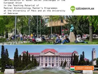
Manifestation of Novel Social Challenges of the European Union in the Teaching Material of Medical Biotechnology Master’s P rogrammes at the University of Pécs and at the University of Debrecen Identification number : TÁMOP-4.1.2-08/1/A-2009-0011.

Regeneration in animal models
E N D
Presentation Transcript
Manifestation of Novel Social Challenges of the European Unionin the Teaching Material ofMedical Biotechnology Master’s Programmesat theUniversity of Pécs and at the University of Debrecen Identificationnumber: TÁMOP-4.1.2-08/1/A-2009-0011
Manifestation of Novel Social Challenges of the European Unionin the Teaching Material ofMedical Biotechnology Master’s Programmesat theUniversity of Pécs and at the University of Debrecen Identification number: TÁMOP-4.1.2-08/1/A-2009-0011 Dr. PéterBalogh and Dr. Péter Engelmann Transdifferentiation and regenerative medicine – Lecture 3 Regenerationinanimalmodels
Regeneration Regeneration is the sequence ofmorphogenetic events that restoresthe normal structure of an organ after its partial or total loss/amputation.Itexistsatdifferentlevelsinplants, invertebrate and vertebrateanimals
Types of regeneration in multicellular organisms Physiological regeneration Reparative regeneration Tissue damage or loss Hypertrophy Morphallaxis
Evolution of stem cells • An ancient conglomerate of flagellate protistsis developed. • All cells at the surface shifted into subgroups of dividing cells and non-dividing cells. Those can be named as unipotent stem cells and non-stem cells. • When multicellularity developed, there was a requirement for migrating stem cells to replace other cells inside the body.
Regeneration in Porifera(sponges) • One cell population of sponges, theso called archeocytes are active stem cells. • Archeocytes are able to differentiate into various types of cells and repopulate themselves by self-renewal. • Archeocytes give rise to choanocytes (participate in respiratory and digestive function), sclerocytes (important cells in innate immunity). • Archeocytesalsoproduce oocytes, while choanocytes produce sperm. • In special occasions choanocytestransdifferentiate into archaeocytes.
RegenerationinHydra • ArtificiallydissectedHydrapolypscanretaintheiraggregationwithin 48 hours. • Individual animals do not increase their size since growth is just balanced by loss of tissue in the form of buds in the lower gastric region and by sloughing of tissue at the ends of the tentacles and from the basal disk.
Stem cell lineages in Hydra • Epithelial cells in the Hydra body column continuously undergo mitotic divisions. Moreover, ectodermal and endodermal epithelial cells also exist. • These two epithelial cell layersmade up by stem cells. Hydra epithelial cells are capable, by successive divisions, both of indefinite self-renewal and of producing different types of specialized cells such as tentacle or foot specific epithelial cells. • In addition, an interstitial stem cell layer is developed.
Molecular factors of Hydra stem cells • Notch signaling • Wnt signaling • BMP molecules • JAK/STAT • Gene screen for stem cell related genes (Sox2+, Nanog, Oct3/4??)
Regeneration in planariaI • Planarians are bilaterally symmetricalanimals found in freshwater streams and ponds. • Planarians have the capacity to replace enormous amount of missing regions through regeneration. • Planarian regeneration is referred toas morphallaxis. • Morphallaxis means cell proliferation / regeneration events away from the wound tissue.
Regeneration in planariaII Neoblasts • Planarian bidirectional regeneration is mediated by neoblasts. • Approx. 30% of the total cells in the planaria are neoblasts • Neoblasts can be found in the entire mesenchymal region of the body with the exception of the pharyngeal region. • Neoblasts divide by mitosis and can repopulate themselves. They are the only dividing cells in planaria. • When a planaria is wounded, neoblasts migrate to the site and begin dividing. • Neoblasts can become any cell type the planaria needs—nerve cells, reproductive cells, etc.
Molecular pattern of neoblasts • Nanos RNA • Piwi RNA • Piwi subfamily - Argonaute proteins • miRNA • Wnt pathway • Shh pathway • FGF family
Regenerative capacity of axons inC. elegans • Axons are able to regrow in many animals after injury except in mammals. • In C. elegansaxonal regeneraton appears as early as 4-5 hoursfollowinglasersurgery, a growth cone-like structure is developed after 6-10 hours. • DLK-1 pathway involved mainly in this regeneration process.
Regeneration in annelids • It hasbeenknown fordecades that annelids are capable for efficient regeneration of their injured body parts. • However the molecular aspects of this regeneration is more hidden. • After 6-10 hours of the injury, neoblast cells are present and give rise to other tissue cells. • Moreover, transdifferentiation of epithelial cells into nerve cells was observed.
Regeneration in insects • Some insects are able to regenerate their legs and other appendage organs. • Other insect species (flies) such as Drosophila do not regenerate adult appendages, but from their imaginal discs they have a great regenerative behaviours during larval life. • In this process several factors are participating such as decapentaplagic (dpp), wingless (wg) etc. molecules.
Regeneration and colony fusion in protochordates • Protochordates share developmental history with vertebrates at least through their early stages. • These colonial metazoans can give hints for regeneration mediated by stem cell activity. • Colony development is dependent on self / non self recognition mediated by a polymorphic gene family (Fu/HC) along with germ and somatic stem cell circulation.
Regeneration of vertebrates • There are two types of regeneration: • Epimorphosis or epimorphic regeneration:This type of regeneration involve the reconstruction of the missing parts by local proliferation from the blastema, or addition of parts to theremaining piece. For example: regeneration of tail, limbs and lens in amphibians and other vertebrates. • Morphallaxisor morphallacticregeneration: This type of regeneration involving reorganization of the remaining part of the body of an animal. For example: Hydra, planaria and other invertebrates e.g. regeneration of the new individual from body pieces.
Regeneration in fish I • Fin regeneration divided into four successive steps: • 1Wound healing / closure within 3 hrs • 2Blastema formation within 1 day • 3Regenerative outgrowth concomitant to differentiation within 2 days • 4Patterning of blastema
Regeneration in fish IIHeterogeneity of cell source • During fin regeneration epidermis contains different compartments of blastema cells: • Distal • Proximal • Lateral
Regeneration in fish IIImolecular patterns • Shh • Wnt • FGF • Activin b A • C-Jun, JunB
Epimorphosis or epimorphic regeneration • Regeneration of tail in amphibians and reptilia: • Amphibia: The tail lacks vertebrae and has an unsegmented cartilaginous tube, which contains the regenerated spinal cord which form mainly of the ependymal lining of the central canal . At first very few cells accumulate under the wound epithelium . The ependyma and the various connective tissues dermis, muscle septa, adipose tissues and osteocytes of vertebrae are the sources of cells for the generate. The non-nervous elements proliferate behind the apex, forming both the muscle and cartilage tube ,then the ependyma proliferate and gradually extend dorsally. • Reptilia: For example lizard, the regenerated tail is a quite imperfect tail. It lacks vertebrae,and in their place, has an unsegmented cartilaginous tube. This tube contains the regenerated spinal cord, including the extension of the ependymal lining of the central canal of the spinal cord.
Similarities in regeneration 1 1 1 1 1 1 2 2 2 2 2 5 PD deletion PD duplication 3 3 3 3 3 4 4 4 4 4 5 5 5 5 2 3 4 Urodele amphibian 5 Intercalation Transplantation Transplantation No intercalation 1 1 1 1 3 2 1 1 1 1 Insect 2 2 2 2 2 3 2 2 2 5 3 3 3 3 3 3 3 3 4 Intercalation Intercalation Transplantation Transplantation 4 4 4 4 4 4 4 5 4 5 5 5 5 5 5
Imprinting in regeneration Amputation Inactivation Off Never expressed Memorized Renewed Activation On On On Gene expression M M M On Off Off Off Off Off A11 A11 A11 On On Off Off Off A13 A13 A13 On On On
Regeneration ofthelimb • Regeneration begins in 3 phases: • 1 Phase ofwound healing or pre -blastema stage: • Blood clotting and migration of epidermal cells from the basal layer of epidermis toward the centre of the wound. The wound is covered with epithelium which is thicker than the epidermis of the limb. • 2Phase of blastema formation: • Cells accumulate beneath the epithelial coverings and form the blastema. Mesenchymal cells accumulate beneath the cap. Mesenchymal – blastemal cells differentiate into myoblasts and muscle cells, early cartilage cells and cartilage. During the dedifferentiation phase hyaluronate (HA) increases in the distal stump to form blastema . As the blastema forms, the HAconcentration will be decreased. The production of HA and break down of collagen represent the establishment of migration from stump tissues. • 3Phase of dedifferentiationand morphogenesis: • The blastema begins to restore the part of which the limb was deprived. Specifically, if the fore arm is removed, the blastema differentiated directly into the muscle, bone, cartilage and skin of the fore arm.
Regeneration ofamphibian lens • 1Afterremovalorinjury of thelensthe dorsal region of the iris thickens and a cleft arises between inner and outer lamellae of the iris. • 2Amoeboid cells move from the stroma into the cleft followed by marked increase of RNA and DNA synthesis as well as of mitotic cell division. • 3 The pigmented cells of the dorsal region is engulfed by invading amoeboid cells. • 4The formed non- pigmented cubical cells form hollow epithelial vesicle and extends with inner and outer lamellae. • 5The vesicle inner wall cells elongated into the lumen and form primary lens fibers. • 6The lens-specific crystalline proteins are formed. • 7The primary lens fibers push to the front of vesicle to form a nucleus behind the lens epithelium which form the secondary lens fibers. • 8The nucleus of primary lens fibers is enclosed by secondary lens fibers. • 9In the central lens fibers the nucleus degenerate ,primary and secondary lens fibers are the components of the lens.
Neuralstemcellsdifferentiationcapacity in vitro Neurogenesis Gliogenesis Noggin Low-RA FGF-2 FGF-2 Passage(6 days) iPScells ES cells Embryoid body Primary neurosphere Secondary neurosphere Blastocysts Differentiation Differentiation Somaticcells Neuron Neuron Astrocyte Oligodendrocyte in vivo Neurogenesis Gliogenesis Blastocysts Embryos Neonatals Adults Earlyneurogenesis Projecting neuron Cholinergic neuronDopaminergicneuronMotor neuron Lateneurogenesis Interneuron GABAergic neuron
Factors controllingregenerationinvertebrates • Nervous system • Animal size • Pituitary gland • Vitamin A and its derivatives • Insulin
Summary • All organisms possess a certain level of regeneration capacity after tissue injury. • At early stage of evolution, animals are able to regenerate whole body, however this capacity is more restricted to specialized tissues and organs in course of evolution. • Neoblasts, hemoblast, progenitor, stem cells are participating in this process.