Hearing
Hearing. Anatomy of the Ear. Ears, Hearing, and Balance

Hearing
E N D
Presentation Transcript
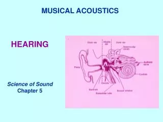
Anatomy of the Ear Ears, Hearing, and Balance • The ears provide the sense of hearing. They also detect head position and motion, so they are essential to balance. The parts concerned with hearing and balance are located in different areas of the ear, but the function of both is based on “hair cell” receptors.
The Outer Ear • The Outer Ear • The outer ear is composed of the ear flap (pinna) and the outer ear canal (external auditory meatus). • The Ear Flap (Pinna) • The pinna collects sound by acting as a funnel • The pinna amplifies the sound and directs it to the auditory canal • Helps to funnel sound waves into the outer ear canal • The filtering effect of the pinna selects sounds in the frequency range of human speech
The Outer Ear • Amplification • Amplification of sound is by the pinna, the tympanic membrane (eardrum) and the middle ear • Frequencies • Low frequencies and high frequencies are treated differently • For low frequencies; it directs sounds toward the ear canal • For high frequencies; they enter the ear canal at a very slight delay • These three parts cause an increase in amplification by about 10 to 15 dB
The Outer Ear Canal The Outer Ear Canal (External Auditory Meatus) • The outer ear canal (external auditory meatus) is slightly S-shaped • The external auditory meatus is a tube that runs from the outer ear to the middle ear. It extends from the pinna to the eardrum and is about 35mm in length and 5 to 10mm in diameter
The Middle Ear • The middle ear is the portion internal to the eardrum. • The mammalian middle ear contains three ossicles. • The primary function of the middle ear is to transfer acoustic energy from compression waves in air to fluid membrane waves within the cochlea.
Ossicles • The middle ear contains three tiny bones known as the ossicles, the maleus, incus and stapes. • They are also referred to as the hammer, anvil, and stirrup. • The malleus and incus evolved from lower and upper jaw bones present in reptiles. • The ossicles are supposed are supposed to mechanically convert the vibrations of the eardrum into amplified pressure waves in the fluid of the cochlea.
Muscles • The movement of the ossicles is controlled by two muscles, the stapedius and the tempor tympani muscle. • The stapedius muscle is the smallest skeletal muscle in the body connects to the stapes and is controlled by the facial nerve. • The tensor tympani muscle connects to the base of the malleus and is controlled by the trigeminal nerve. • These muscles contract in response to loud noises, reducing the transmission to the inner ear, this is known as the acoustic reflex.
The Inner Ear The Inner Ear • Composed of the cochlea, semicircular canals, and vestibule, which are all linked together • The semicircular canals have three canals; the posterior, the superior, and the lateral • All are filled with fluid and all are protected by the temporal bone
Hair Cells Hair Cells • Each hair cell is seen to have 40 to 100 hairs arranged in a curve • Nerve fibers run from the cell bases
The Process of Balance • A process involving a range of sensory inputs, analysis in the brain and motor outputs • Inputs arrive from the eyes, micro-receptors in muscle tendons, and skin pressure sensors • The inner ear’s fluid-filled vestibule and semicircular canals play a key role • They incorporate sensitive hair cells similar to the cochlea’s • The gravity (static equilibrium) react to the speed and direction of head movements (dynamic equilibrium) • Both equilibriums respond to most head positions and movements
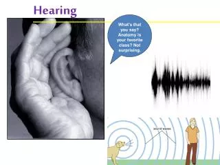
How we Hear When we hear noises, our body is converting energy from sound waves into nerve impulses which are interpreted by the brain. Sound waves are produced when air is mechanically disturbed. Sound is measured in two different ways: • Frequency: • also known as the pitch (high or low) • Frequency is measured by the number of complete sound waves per second • High pitch sounds can be damaging to hearing. 2. Intensity: • Loudness
How we Hear What happens when we hear a noise… • Sound waves enter the ear canal, and make the ear drum vibrate • Vibrations travel through the 3 connected bones located in the middle ear • This motion forces fluid movement in the inner ear • The movement of the fluid bends thousands of tiny, delicate hair-like cells. This process converts vibrations into nerve impulses
How we Hear 5. Nerve impulses are transported to the brain via the auditory nerve 6. Finally, the impulses are converted into what we hear as sound http://www.sc.edu/ehs/modules/Noise/hearing.htm
Hearing Loss • the ear is a delicate structure • makes it easy for the ear to get damaged • when the eardrum is damaged or the tiny bones of the middle ear lose their ability to vibrate, the ear loses its ability to conduct vibrations
Hearing Loss • if damage occurs to the cochlea’s hair cell receptors or their nerves, the damage can cause sensorineural hearing loss or nerve deafness • diseases sometimes cause sensorineural hearing loss • the main reasons for sensorineural hearing loss are biological changes such as heredity, aging, and exposure to loud music or noise
Hearing Loss • when the tissues are destroyed they remain dead • using a hearing aid may develop enough sound to stimulate neighboring hair cells • when digital hearing aids are used • they improve hearing by: • amplifying vibrations for frequencies • compressing sound
Hearing Loss • a cochlear implantis a sort of bionic ear • it is the only way to repair hearing for those with nerve deafness • a cochlear implant translates sounds into electric signals • these signals carry information about the sound to the brain