Download
1 / 15
150 likes | 361 Vues
REVERSIBILITY OF MORPHOLOGICAL CHANGES IN THE MESENTERIC LYMPH NODES AFTER PERORAL ADMINISTRATION OF GOLD NANOPARTICLES Olga V. Zlobina, Saratov State Medical University , Russia Svetlana S. Pakhomy, Saratov State Medical University , Russia

E N D
REVERSIBILITY OF MORPHOLOGICAL CHANGES IN THE MESENTERIC LYMPH NODES AFTER PERORAL ADMINISTRATION OF GOLD NANOPARTICLES Olga V. Zlobina, Saratov State Medical University , Russia Svetlana S. Pakhomy, Saratov State Medical University , Russia Alla B. Bucharskaya, Saratov State Medical University , Russia Irina O. Bugaeva, Saratov State Medical University , Russia Galina N. Maslyakova, Saratov State Medical University , Russia Nikolai G. Khlebtsov, IBPPM RAS, Russia Boris N. Khlebtsov, IBPPM RAS, Russia Vladimir A. Bogatyrev, IBPPM RAS, Russia
The purpose of the study To study the severity and reversibility of structural and functional changes in the mesenteric lymph nodes of rats with regard to the size and duration of oral administration of gold nanoparticles Reactive center of cortical substance of lymphoid follicle (x1000)
Objectives 1. To study the effect of the size of orally administered gold nanoparticles on the cellular composition of the mesenteric lymph nodes 2. To analyze the recovery rate of the initial cellular composition of mesenteric lymph nodes after cessation of administration of gold particles of different sizes 3. Determine the recovery time the initial cellular composition of mesenteric lymph nodes after oral administration of gold nanoparticles
Materials and methods 48 mature outbred male rats weighing 180-220 g (12 animals per group) Gold nanoparticles size of 1-3, 15, 50 nm (IBPPM RAS, Saratov) 3 experimental groups (oral administration of gold nanoparticle with size of 1-3, 15, 50 nm for 8 days ) Control group (oral administration 1 ml of saline for 8 days ) The lymph node biopsies were taken for histological examination a day after cessation of oral administration (6 animals per group) The lymph node biopsies were taken for histological examination 14 days after cessation of oral administration (6 animals per group)
Methods of study of histological preparations mesenteric lymph nodes • Hematoxylin-eosin (for counting cell components). • Toluidine blue at pH Vinogradov - 4.8 • (to identify metachromatic mast cells). • 3. Methyl green pyronine by Brachet • (for the detection of DNA and RNA). • 4. Smears, painting Romanovsky-Giemsa.
Methods of study of histological preparations mesenteric lymph nodes Computer system to control the size of the micro-and nanometer-scale structures Image Analyzer. The standard method of counting the cell elements at a magnification of 200, 400, 900, 1350 using specialized morphometric grid (Avtandilov GG, 1972, 1992). Morphometric image analysis program - ImageJ.
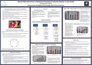
Results: a day after administration 1. The reactions of the cellular elements of the mesenteric lymph nodes a day after introduction of nanoparticles were size dependent. 2. The statistically significant dynamics was not observed in the group treated with gold nanoparticles of 1-3 nm. 3. The introduction of nanoparticles of 15 nm and 50 nm led to significant changes in the content of the cellular elements of the lymph nodes and was more reflected in the paracortical zone than in areas of lymphoid follicles and brain strands.
Results: a day after administration • The presence of nanoparticles in animal organism intensified the process of blast transformation of lymphocytes • The control group - immunoblasts were absent in paracortical area of lymph nodes; their number increased to at administration of 15 nm and 50 nm nanoparticles (to 2,6 ± 0,4 and 2,9 ± 0,1 respectively) . • Mast cells present only in the area of brain strands in control and experimental groups after introduction of nanoparticles, whose number increases in direct proportion to the size of the nanoparticles. • In all the studied areas of lymph nodes the increased frequency of mitotic figures was noted after nanoparticle administration.
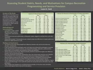
Control Zone of lymph.follicles Paracortical zone Zone of brain cords 1-3 nm 15 nm 50 nm Changes in the number of small lymphocytes after 8 day introduction of nanoparticles of different sizes
50 nm 15 nm 1-3 nm control The changing of immunoblasts number in zone of lymphoid follicles after oral administration of nanoparticles
Results: a day after administration b a Lymph node after nanoparticle administration (a) 1-3 nm; (b) 15 nm (х400). The data indicate the marked immune modulating effect of gold nanoparticles in animals, depending on the nanoparticle size and organ specificity. A pilot study revealed a certain temporal dynamics of cellular responses.
Results - the reversibility of the morphological changes • The cellular composition of the lymph nodes after the introduction of gold nanoparticle size of 1-3 nm for 8 days corresponded the control values • The number of lymphocytes and immunoblasts decreased less significant and was not reaching the control values after cessation of 15 nm nanoparticles administration. It may be due to incomplete restoration of the original state of the immune system 14 days after cessation of nanoparticle administration
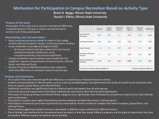
Regression analysis of the recovery rate of the cellular composition of lymph nodes after cessation of 15 nm nanoparticle administration
Regression analysis of the recovery rate of the cellular composition of lymph nodes after cessation of 15 nm nanoparticle administration 1. Duration of initial recovery of cellular composition of the lymph nodes does not exceed 21 days, in connection with which it can be concluded that 15 nm nanoparticles have not immunotoxicity actions at 8 days oral administration 2. 14 days after cessation of administration of 50 nm nanoparticles it may be noted that cellular composition did not corresponds to the control values. By regression analysis it was found that 21-st day is predicted to achieve the control values of cellular composition.
Conclusions 1. Oral administration of gold nanoparticles leads to changes in the cellular composition of the mesenteric lymph nodes, the severity and reversibility of the reaction is determined by the size of nanoparticles 2. Administration of gold nanoparticles with size 15 nm and 50 nm leads to a change in the cellular composition of nodes on the 8th day. The number of medium and large lymphocytes are almost doubles in brain cords, the content of plasma cells is increasing, degranulation signs of mast cells are appearing in subcapsular zone 3. 14 days after cessation of 1-3 nm gold nanoparticle administration the number of small and medium lymphocytes, plasma cells, mast cells, and mitotic figures corresponds to the initial values. According to a regression analysis the number of immunoblasts and large lymphocytes corresponds to the control values after 16 days, indicating the absence of immunotoxicity.