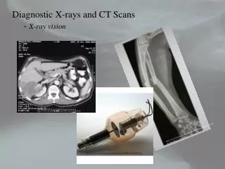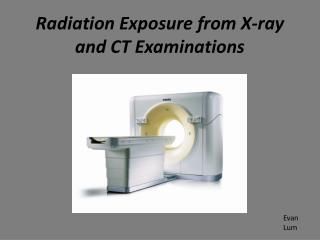
CT Instrumentation and X-ray system
250 likes | 692 Vues
CT Instrumentation and X-ray system . Alhanouf Alshedi Email: aalshedi@ksu.edu.sa. 2 ed Lecture. Scanner. GANTRY PATIENT COUCH . Gantry :. Is a mounted framework that surrounds the patient that houses these components: X-ray tube Generator Collimators

CT Instrumentation and X-ray system
E N D
Presentation Transcript
CT Instrumentation and X-ray system Alhanouf Alshedi Email: aalshedi@ksu.edu.sa 2ed Lecture
Scanner GANTRY PATIENT COUCH
Gantry : Is a mounted framework that surrounds the patient that houses these components: X-ray tube Generator Collimators Detectors Filters
X-ray tube: • Uses high frequency generator. • To maximize x-ray tube heat capacity: • Uses rotating anode x-ray tube. • small target angle. • large anode diameter • focal spot size appropriate to geometry • Some use a metal envelope, which have larger anode disks. This allows the tech. To use higher tube current and heat capacity is also increased. • Cathode consists of one or multiple filaments.
mA – tube current: The number of electrons flowing from cathode to anode. kVp: Potential difference between cathode and anode (Volts) kilo means 1,000 x. S –time of exposure:mAstube current for certain length of time.
Generator High –frequency generator which is : • Small • Compact • More efficient than conventional generators. • Is located inside the gantry. • Provides power ranging 20-100 kilowatts
Filters: They serve a dual purpose: • Filtration removes long-wavelength x-rays as do not play a role in C.T image formation and add to patient dose. • Filtration shapes the energy distribution across the beam to produce a uniform beam.
- Adjustable pre-patient (between tube & patient) and pre-detector collimators: (between patient and detector) • - Must be perfectly aligned to optimise imaging process. Collimation: • It protects the pt by restricting the beam to area of interest. • Shape the beam and removes scatter radiation which improves axial resolution.
Tube Detector Pre-Collimation Pre-collimator • Constrains size of beam. • Reduces amount of scatter produced. • Designed to minimize beam divergence. • Often consists of several stages or sets of jaws.
Tube Detector Post-Collimation Post-collimator Helps define slice (beam) thickness. Reduces scatter radiation reaching detector
GANTRY CHARACTERISTICS Has two important features: 1)APERTURE: It is the opening in which the pt moves through during scanning. Most of the scanners have 70cm aperture, which facilitates pt positioning and provides access to pt in emergency situations. 2)TILTING RANGE: To accommodate all patients and different clinical exams. Tilting range of most scanners +30 to -30 degrees.
PATIENT COUCH Should be strong and rigid to support weight . Usually made of carbon fibers due to their low absorption. 450 lbs (204 kg) distributed weight limit. Provides vertical and horizontal movement. Scannable range:coverage from head to thigh (162cm)
Any Question? Thank You