Download

1 / 56
810 likes | 1.87k Vues
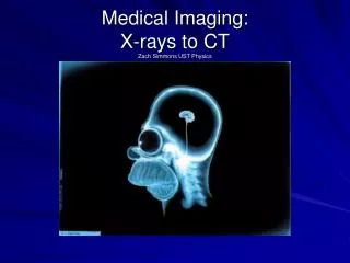
Course 046831. Introduction to Medical Imaging X-ray and CT. Guy Gilboa. X-Ray. First imaging modality (Discovered by Röntgen in 1895). X-ray discovery. Wilhelm Rontgen 1895 - Discovered and detected X-rays Used it as a first medical imaging modality.

E N D
Course 046831 Introduction to Medical Imaging X-ray and CT Guy Gilboa
X-Ray First imaging modality (Discovered by Röntgenin 1895).
X-ray discovery • Wilhelm Rontgen • 1895 - Discovered and detected X-rays • Used it as a first medical imaging modality. • Nobel 1901, first Noble prize in physics was awarded to him. First x-ray image. Hand of Anna Rontgen (wife)
X-ray machines Standard machine C-arm
X-ray tube diagram Taken from http://used-medicalequipmentblog.blogspot.co.il/2011/05/advances-in-ct-scanner-x-ray-tubes.html
Energy units eV = electron- Volt It is equal to the amount of kinetic energy gained by a single unbound electron when it accelerates through an electric potential difference of one volt. Photon energy: kVp= peak kilo-voltage. The maximum voltage applied across an X-ray tube. Determines the kinetic energy of the electrons accelerated in the X-ray tube and the peak energy of the X-ray emission spectrum. h – Planck’s constant - wavelength c – speed of light
Standard optics constraints affect the quality of the X-ray image Taken from [1] http://www.medphysics.wisc.edu/~block/bme_530_lectures.html
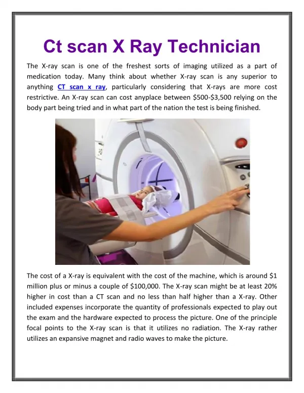
Popular for checking chest (lung problems) and bone fractures
Signal to Noise Ratio • Poisson distribution • Variance is • SNR
X-Ray Summary Advantages: • Cheap and simple • Low radiation (compared to CT) Drawbacks: • Does not give 3D info. • Bones can occlude significant diagnostic data.
Digital Mummography • Used to detect small tumors or microcalcifications in the breast. • Very high spatial resolution can CNR are need for these type of pathologies (often <1mm in diameter) • Low radiation is important – avoid tissue damage and allow frequent usage. • Low energy (e.g. 26 keV) is used – high contrast, low radiation, low penetration.
Computed Tomography (CT) 3D imaging using X-ray radiography
History - Invention of CT • Sir Godfrey Hounsfield (English Electrical Engineer), built first CT 1971, scanned head. • Allan McLeod Cormack - math framework. • Nobel prize for both in 1979 for the invention of CT. • 1975 – first full body scanner. Hounsfield sketch
CT – operation principle Taken from http://www.cyberphysics.co.uk/topics/medical/CTScanner.htm
Attenuation coefficients • Linear attenuation coefficient • N – number of X-rays transmitted through a certain thickness x of tissue
Hounsfield unit Water: 0HU Air: -1000HU What is displayed in CT images? Typical medical scanner display: [-1024HU,+3071HU], 12 bit per pixel is required in display. Range:
Hounsfield units of tissues Taken from [1]
Rendering based on different HU thresholds Taken from https://www.sharbor.com/news/MSG/index.html
X-ray detectors • Previously – film, analog radiography. • Today – digital radiography • Indirect conversion • X-ray to light using a scintillator (CsI:T1) • Light to voltage using photodiodes • Direct conversion • Thin-Film-Transistors • Cadium-Teloride, Cadium-Zinc-Teloride – technology not mature yet.
Helical CT Taken from [1]
Scanning Geometry of a CT System From [1]
Gantry rotation speed (times) • Range • Standard 0.4 – 0.6 sec/rot • Cardiac 0.27 – 0.35 sec/rot Gantry rotation: https://www.youtube.com/watch?v=YqOSBJO0_-g
Multi Slice Detectors Taken from http://tech.snmjournals.org/content/36/2/57/F1.expansion.html
Reconstruction • Collimators are used to keep the exposure to a slice. • Image is built from multiple projections. • Parallel rays are often assumed – simplifies the math. Preprocessing is done for fan-beam – conversion to parallel structure.
Radon transform • Input space coordinates x, y • Input function f(x, y) • Output space coordinates a, s • Output function F(a, s) 2D Geometry Taken from http://uprt.vscht.cz/prochazka/pedag/
Transform and Its Inverse There are several possible parameterizations of the transform, here is one: Radon transform Inverse Radon transform
Filtered Back Projection The common fast and robust way to reconstruct
FBP illustration From [1] And another illustration https://www.youtube.com/watch?v=BhOMbjXzjP8
Iterative Reconstruction • A more sophisticated way to reconstruct. • Can incorporate very accurate modelling of the physical projections. • Estimates an initial solution and iteratively forward and backward projects until convergence. • Slow, (considered state-of-the-art). Taken from http://www2.alasbimnjournal.cl/alasbimn/CDA/imprime/0,1208,PRT%253D455,00.html
Fan beam reconstruction Measurements are rearranged to form a parallel geometry representation.
Helical CT Allows continuous gantry rotation – z axis is changing continuously, this is taken into account in the reconstruction.
Contrast agents • Iodine is injected – increase attenuation coefficient of blood for a short time. liver vessels
Dual energy CT • Can distinguish better between materials with different attenuation coefficients at different energies.
Dual energy CT – several methods • Dual source (Siemens)
Dual energy CT • kV Switching (GE)
Dual energy CT • Dual layer (Philips)
Spectral CT by photon counting • Future technology using direct conversion methods. • In 2014, not a mature technology yet (works for animals, mummograph).
Spectral CT - example CT image of the thorax of a mouse injected with Au-HDL and iodine contrast agents. Conventional vs. photon-counting CT. Taken from http://www.medical.philips.com/us_en/about/News/Publications/MedicaMundi/Cormode-Fayad.wpd
Uses of CT Used widely to scan almost every organ in the body, popular uses are: • Cerebral scans – chronic and accute head and brain scans, internal bleeding, tissue oedema (swelling) and skull fracture. Also to diagnose and follow the progression of some brain tumors. • Pulmonary disease – identify size and geometry of lesions. Calcification of nodules, increase in tissue attenuation – can be indicators for cancer.
CT Uses (cont’) • Liver imaging - 2-phase liver scan: • A pre-contrasted control scan is acquired as baseline • Two scans following injection of contrast after ~35sec (arterial phase) and ~65sec (portal phase). • Can detect hypervascularlessions, fatty infaltrations into the liver and other liver problems. • Cardiac imaging • Used primarily for assessing calcifications within the heart, particularly in coronary arteries. • The presence of coronary calcifications is highly predictive of the future development of cardiac problems.
CT Uses (cont’) • (Cardiac – cont’) • Iodine contrast is used. High end machines are needed – with fast gantry rotation to “freeze” the cardiac motion. • Can work with pacemakers, defibrillators, stents etc. where MRI cannot work. • Trauma– as CT becomes available and fast – it is commonly used in trauma (ER) – a full body scan is often performed to diagnose internal bleeding and bone fractures.