General introduction- fundamental principles X ray & CT
530 likes | 805 Vues
General introduction- fundamental principles X ray & CT. Radiology Dept. 1st Hospi. of Peking University 唐光健. Questions to think of with the lecture. What is X ray? What kinds of the features of it are useful or harmful? What are the two steps of X ray imaging?

General introduction- fundamental principles X ray & CT
E N D
Presentation Transcript
General introduction- fundamental principles X ray & CT Radiology Dept. 1st Hospi. of Peking University 唐光健
Questions to think of with the lecture • What is X ray? What kinds of the features of it are useful or harmful? • What are the two steps of X ray imaging? • How is the grey scale of bone, muscle and air in the X ray film? • What is the main difference between the CT imaging and the image of X ray film? • What are same and what are different between the processes of CT imaging and imaging of X ray projection? • Is the meaning of a CT axial imaging and a section of a body specimen in same location identical?
Black box? Wilhelm C Röntgen
Generation and features of X ray • High speeding electrons Anode target sudden slow down special X ray – sequence spectrum narrow consecutive X ray – sequence spectrum wide e special X ray – sequence spectrum narrow consecutive X ray – sequence spectrum wide e X ray
Generation and features of X ray • electromagnetic waves -ionizing radiation -wave length of medical use: 0.08 ~ 0.71Å -feature of wave; feature of particle • Features-Effects • Physical effect • Chemical effect • Biotic effect
Generation and features of X ray • electromagnetic waves -ionizing radiation -wave length of medical use: 0.08 ~ 0.71Å -feature of wave; feature of particle • Features-Effects • Penetrability • Fluorescent effect • Thermal effect • Ionizing effect • interference,diffraction, reflection, refraction • Physical effect • Chemical effect • Biotic effect
Generation and features of X ray • Penetrability • Fluorescent effect • Thermal effect • Ionizing effect • interference,diffraction, reflection, refraction • Features-Effects • Physical effect • Chemical effect • Biotic effect -be able to penetrate ma. of dif. density while attenuated I = I0 · e-µl I0 µ I l
Generation and features of X ray • Penetrability • Fluorescent effect • Thermal effect • Ionizing effect • interference,diffraction, reflection, refraction • Features-Effects • Physical effect • Chemical effect • Biotic effect -making some compounds fluoresce -Fluoroscopy, image intensifier
Generation and features of X ray • Penetrability • Fluorescent effect • Thermal effect • Ionizing effect • interference,diffraction, reflection, refraction • Features-Effects • Physical effect • Chemical effect • Biotic effect -making some compounds fluoresce -Fluoroscopy, image intensifier
Generation and features of X ray • Penetrability • Fluorescent effect • Thermal effect • Ionizing effect • interference,diffraction, reflection, refraction • Features-Effects • Physical effect • Chemical effect • Biotic effect -Subject absorb the energy of X ray ionized ionized voltage -ionizing cell auto-exposure X ray detector
Generation and features of X ray • Penetrability • Fluorescent effect • Thermal effect • Ionizing effect • interference,diffraction, reflection, refraction • Features-Effects • Physical effect • Chemical effect • Biotic effect - penumbra, interferential signal image blued
Generation and features of X ray • sensitization effect • coloration effect fundament of X ray filming • Features-Effects • Physical effect • Chemical effect • Biotic effect
Generation and features of X ray • Features-Effects • Physical effect • Chemical effect • Biotic effect • radiated tissue cells restrained, damaged, necrosing X ray protection
X ray Imaging • X ray penetrate the subject(body) while attenuated • attenuate signal recorded by the accepting medium
X ray Imaging • X ray penetrate the subject(body) while attenuated • attenuate signal recorded by the accepting medium I0 µ1 µ2 µ3 µ4 I l1 l2 l3 l4 I = I0 · e-µ1l1· e-µ2l2· e-µ3l3· e-µ4l4
X ray Imaging • X ray penetrate the subject(body) while attenuated • attenuate signal recorded by the accepting medium • getting special distribution of different density overlapping of anterio-posterior images
X ray Imaging • X ray penetrate the subject(body) while attenuated • attenuate signal recorded by the accepting medium • film-silver bromide decomposed positive correlation with dosage of X ray negative correlation with degree of attenuation • flu. screen • detectors
X ray Imaging • X ray penetrate the subject(body) while attenuated • attenuate signal recorded by the accepting medium • grey of spots ∽ thickness & density of the mat. in the path way of X ray beam • distribution of the spots ∽ anatomic form in the path way of X ray beam • difference between the sports - contrastimage natural contrast artificial contrast
X ray Imaging • X ray penetrate the subject(body) while attenuated • attenuate signal recorded by the accepting medium • grey of spots ∽ thickness & density of the mat. in the path way of X ray beam • distribution of the spots ∽ anatomic form in the path way of X ray beam • difference between the sports - contrastimage natural contrast artificial contrast
X ray Imaging • X ray penetrate the subject(body) while attenuated • attenuate signal recorded by the accepting medium • grey of spots ∽ thickness & density of the mat. in the path way of X ray beam • distribution of the spots ∽ anatomic form in the path way of X ray beam • difference between the sports - contrastimage natural contrast artificial contrast
X ray equipment • X ray source • X ray tube High voltage generator • X ray imaging devices • X ray film / image intensifier / plate detector • X ray mechanical device • exam table frame……
X ray equipment • X ray tube • filament - cathode • anode target • tube shell
X ray equipment • X ray tube • filament - cathode • anode target • tube shell
Protection from X ray • X ray protection • Cancel X ray exam unnecessary • Reduce radiation dosage • Distance from X ray source • Protective device
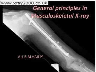
Principles of imaging diagnosis • familiar with imaging methods and body position • familiar with the normal imaging anatomy and variations • sequential observation • imaging manifestations and relationship with neighboring structures of the lesion • clinical and laboratory materials • understanding the meaning and limitations of the imaging methods
fundamental principles of CT • CT-Computerized Tomography • real tomography,without any overlapping
fundamental principles of CT I0 I0 µ1 µ1 µ2 µ3 µ3 µ4 I I µ2 µ4 l1 l1 l2 l2 l3 l3 l4 l4 l1 = l1 = l1 = l1 = 1 I = I0 · e-µ1l1· e-µ2l2· e-µ3l3· e-µ4l4 I = I0 · e-µ1l1· e-µ2l2· e-µ3l3· e-µ4l4 I = I0 · e- (µ1+ µ2 + µ3 + µ 4) ·1
fundamental princ. of CT First laboratory CT scanner 1968 Godfrey Hounsfield
fundamental princ. of CT First clinical head CT scannerAtkinson Morley‘hosp.,London
fundamental principles of CT • measurement
fundamental principles of CT DAS-data acquisition system -detector -bumper -integrator -amplifier -A/D converter • measurement detector
fundamental principles of CT • Reconstruction
fundamental principles of CT • Reconstruction • backprojective method • Iterative method • Factorial method • Fourier method • Filt. backprojective method
fundamental principles of CT • Reconstruction • Filt. function(Kernol) • High filt.(bone algorithm)low filt.(soft tissue algorithm)
fundamental principles of CT • Reconstruction • Filt. function(Kernol) • High filt.(bone algorithm)low filt.(soft tissue algorithm)
fundamental principles of CT CT value - relative magnitude to water may be used qualitatively but not quantitively • Reconstruction [(μobj.- μwater)/μwater]×1000
fundamental principles of CT • image display
fundamental principles of CT • image display 200/30=7HU muscle 50HU- fat -50HU=100 ~13 grey scale 1000/30=33HU muscle 50HU- fat -50HU=100 ~3 gray scale • ability of identify grey scale • eyes:24~30 • CT:2000
fundamental principles of CT • image display • Voxel and pixel • voxel – 3D, pixel – 2D • CT value of a voxel represent all messages of the elements in the voxel
fundamental principles of CT • image display partial volume effect
fundamental principles of CT • enhancement CThigh density contrast agent injected into veinintro-vascular/-tissue(extracellular space) CT scan density contrast • hypersensitiveness nephrotoxitity
fundamental principles of CT • enhancement CThigh density contrast agent injected into veinintro-vascular/-tissue(extracellular space) CT scan density contrast • hypersensitiveness nephrotoxitity
fundamental principles of CT • enhancement CThigh density contrast agent injected into veinintro-vascular/-tissue(extracellular space) CT scan density contrast • hypersensitiveness nephrotoxitity
fundamental principles of CT • Indications • No absolute contraindication, suitable for emergency and serious cases exam • Single imaging factor,image interpretation easily,anatomy clear and detailed • deformation、infection、trauma、neoplasm… • Limitations • Low soft tissue resolution ,difficult to detect lesion with small density difference from neighbor structure • Low sensitivity for the lesion with slight gross change • Contrast agent using