Download
1 / 17
190 likes | 790 Vues
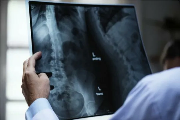
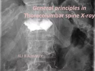
General principles in Thoracolumbar spine X-ray. ALI B ALHAILIY. background. Each year, more than 150,000 persons in North America sustain fractures of the vertebral column. Injuries to the thoracolumbar and lumbar spine constitute most of these fractures. anatomy. L SPINE Trauma.

E N D
General principles in Thoracolumbar spine X-ray ALI B ALHAILIY
background • Each year, more than 150,000 persons in North America sustain fractures of the vertebral column. • Injuries to the thoracolumbar and lumbar spine constitute most of these fractures.
L SPINE Trauma • The lumbar vertebrae are the 5 largest and strongest of all vertebrae in the spine. • These vertebrae comprise the lower back. • They begin at the start of the lumbar curve (ie, the thoracolumbar junction) and extend to the sacrum. • The strongest stabilizing muscles of the spine attach to the lumbar vertebrae. • Fractures of lumbar vertebrae, therefore, occur in the setting of either severe trauma or pathologic weakening of the bone.
Osteoporosis • Osteoporosis is a progressive bone disease that is characterized by a decrease in bone mass and density which can lead to an increased risk of fracture. In osteoporosis, the bone mineral density (BMD) is reduced and the amount and variety of proteins in bone are altered. • Osteoporosis is the underlying cause of many lumbar fractures, especially in postmenopausal women. Osteoporotic spinal fractures are unique in that they may occur without apparent trauma. However, a thorough diagnostic workup is always required to rule out spinal malignancy.
Fracture of the lumbar spine can occur whenever forces applied to the lower spinal column exceed the strength and stability of the spinal column unit . • Common injuries resulting in fractures of the lumbar spine include • fall from a height • motor vehicle and motor vehicle • pedestrian accidents • penetrating trauma, including gunshot wounds and stabbings. • Fractures of the pelvis often are associated with injury to the sacral plexus and the lower lumbar spine.
treatment • The treatment of lumbar compression fractures that are secondary to osteoporotic bone changes has increasingly emphasized the role of vertebroplasty. • Vertebroplasty and kyphoplasty are similar medical spinal procedures in which bone cement is injected through a small hole in the skin into a fractured vertebra with the goal of relieving back pain caused by vertebral compression fractures. • The outcomes of such treatment are helpful in the management of pain.
Postsurgical imaging • Postsurgical imaging must consider the appearance and the position of posterior pedicle screws, which are often used to stabilize lumbar spinal fractures. The long-term value of posterior spinal fixation compared with other means of stabilization remains controversial. • Malposition of pedicle screws and the mechanical failure of pedicle screws have been reported.
Thoracolumbar spine Imaging • Incorrect management of patients with spinal injury may lead to, or exacerbate, neurological deficit. Therefore patients with suspected spinal injury should be managed by experienced clinicians in accordance with local and national clinical guidelines. Imaging should not delay resuscitation. • Further imaging with CT or MRI (not discussed) is often appropriate in the context of a high risk injury, neurological deficit, limited clinical examination, or where there are unclear X-ray findings.
Good views of the T-spine and L-spine are difficult to achieve in the context of trauma. Clinical assessment is also often limited by distracting injuries or reduced consciousness. The clinico-radiological assessment of suspected T-spine or L-spine injuries therefore depends on careful consideration of both the clinical and radiological findings.
Thoracolumbar spine X-ray - General principles • Please have a look on the systemic approach and viewing principles tutorials at: • http://radiologymasterclass.co.uk/tutorials/musculoskeletal/x-ray_trauma_spinal/x-ray_thoracolumbar_spine_normal.html