Download
1 / 25
250 likes | 293 Vues
Explore the properties, hazards, and protection methods related to X-ray technology. Learn how X-rays work, their impact on operators and patients, and essential safety precautions. Discover key components of an X-ray room and the modern X-ray tube. Observations and demonstrations illustrate the principles behind X-ray technology.

E N D
X-ray Principles & Physics Laboratory Russell L. Wilson , CRT, RT(R)
Properties of X-ray • X-rays travel in a straight line and diverge from their point of origin. • X-ray photons have many different energies. • X-rays are highly penetrating. • X-rays are invisible. • X-rays travel at the speed of light.
Properties of X-ray • X-rays produce scatter radiation when they enter-act with matter. • X-rays affect radiographic and photographic film. • X-rays cause fluorescence of some materials. • X-rays cause biologic damage.
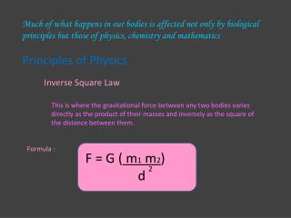
Properties of X-ray • X-rays respond according to the inverse square law.
X-ray Protection • Because x-rays cause biologic damage, the operator of the machine and the patient must be protected from the radiation. • Lead is used to absorb radiation.
X-ray Hazards • In the early days of radiography, the patient often was burned by the radiation. • With proper operation of equipment, x-rays are relatively safe today.
X-ray Injuries Still Occur • These are serial photographs on a patient that had multiple long fluoroscopic examination. • Last image is after skin graphs.
Patient Radiation Protection • Later in the quarter, we will covers methods used to keep the exposure to the patient as low as possible. • Keeping the exposure low is the responsibility of the operator of the x-ray machine.
X-ray Hazards • With early x-ray machine, there was a real possibility of electrocution. • Today with proper safety precautions, radiography is very safe for the operator.
X-ray Hazards • X-ray was also very hazardous for the operator in those early years. • X-ray operators would use their hands to make sure the machine was working .
X-ray Hazards • It was not uncommon for both the operator and patient to receive burns. • Today, with proper precautions, x-ray is safe for the operator.
The X-ray Room • The radiographic equipment consists of: • The Tube Stand, Tube & Collimator • Grid Holder or Bucky • Controls in the Control Booth.
The X-ray Room • The wall of the x-ray room and door are shielded with lead to protect the operator and staff.
Operator X-ray Protection • The Door to the X-ray Room contains lead. • It must be closed during exposures.
Operator X-ray Protection • The wall of the control booth is leaded. Stand completely behind the wall during exposures.
Operator X-ray Protection • Observe the patient or experiment through the lead glass window. No peeking around the wall!
X-ray Tube • X-rays are produced inside the x-ray tube. • Many properties of light and x-ray are the same.
Modern X-ray Tube • This is a modern rotating anode general • radiographic x-ray tube. • The leaded glass holds the vacuum in the tube. • Anode rotated to cool tube.
X-ray Collimator • Using light in the collimator, lead shutters are moved to restrict the area of exposure.
X-ray Collimator • Collimation is our best tool for reducing radiation exposure to the patient.
Observations • 1. Did the light field match the x-ray beam? Yes • 2. What principle did this demonstrate? X-ray s travel in a straight line line and diverge from the point of origin. X-rays have similar properties to light.
Observations • 3. Did the intensity of the fluorescence of the screen change when the kVp was increased? Yes • 4. Would this indicate that the intensity of the beam changed? Yes • 5. During the exposure could you see inside the phantom? Yes
Observations • 6. What property of x-ray did this demonstrate? X-rays are highly penetrating. • 7.After the tone from the control terminated, did the screen continue to fluoresce? No • 8.If the screen did not fluoresce, was there any radiation coming from the tube after the tone stopped? No
Observations • 9. Did you hear any noise coming from the tube after the tone stopped? Yes the rotor continued to rotate. • 10. What did you see on the film that was sitting near the phantom? A blurry image. • 11. What principle did this demonstrate? X-rays produce scatter radiation. Scatter radiation is not divergent.
The End Return to Lectures Home Page