Download

1 / 28
980 likes | 2.49k Vues
Introduction to Medical Imaging. Jeff Benseler, D.O. Objectives. Medical Imaging: What to expect in your first 2 years at OUHCOM Overview: How do x-rays create an image of internal body structures? What are the advantages of CT, MRI and Ultrasound?. Medical Imaging (Radiology).

E N D
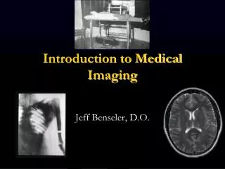
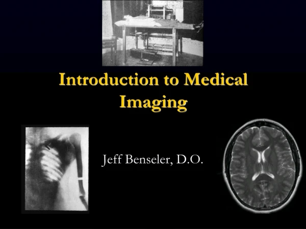
Introduction to Medical Imaging Jeff Benseler, D.O.
Objectives • Medical Imaging: What to expect in your first 2 years at OUHCOM • Overview: How do x-rays create an image of internal body structures? • What are the advantages of CT, MRI and Ultrasound?
Medical Imaging (Radiology) • Nearly all medical disciplines utilize medical imaging • As you move from block to block we will learn: • How each modality works to create an image of internal body structures • Selecting the best imaging tests for a given clinical presentation • Develop a stepwise repeatable pattern of evaluating medical images
Method for learning medical image interpretation • Most blocks will contain recorded presentations • These recordings last approximately 10 to 30 minutes each • Most blocks will have 2 to 4 recordings to view before the live class • The recordings can be viewed and reviewed as needed anytime 24/7 • In class, we will learn by interpreting unknown cases
Questions about medical imaging • Please feel free to contact me with questions • My preferred contact method is email • benseler@ohio.edu
Objective 2 What are x-rays? • No mass • No charge • Energy X-rays are a type of electromagnetic energy
How do x-rays passing through the body create an image? • X-rays that pass through the body render the image dark (black) • X-rays that are totally blocked render the image light (white) • Air = low atomic # = x-rays get through = image is dark (black) • Metal = high atomic # = x-rays blocked = image is light (white)
5 Basic Radiographic Densities 1. • Air • Fat • Soft tissue/fluid • Mineral • Metal 4. 5. 2. 3. Name these radiographic densities.
Optimal environment for visual perception • Dedicated source of light (5 to 9 mega pixel monitors) • Darkened environment (like a movie theater) • Limit distraction
Can you recognize shapes and density?
Find the pathology What clues do you have?
Medical Imaging Interpretation3 basic steps • First learn how each modality creates an image of internal body structures • Next, be able to accurately label normal anatomy (body structures) • Then, search for structures that don’t belong and for body structures that are abnormal in size, shape, position and/or density
History: 11 year old twisting injury of the foot
Naming the parts of a long bone Distal 3. 2. 1. Proximal Word bank: epiphysis, metaphysis, diaphysis, cortex, medullary cavity
Review: What are the 5 basic radiographic densities from black to bright white? Air Fat Soft tissue/fluid Bone/mineral Metal
Summary for objective 2: How do x-rays create an image of internal body structures? • X-rays pass through the body to varying degrees • Higher atomic number structures block x-rays better, example bone • Lower atomic number structures allow x-rays to pass through, example: air in the lungs
Objective 3Advantages of CT, MRI and Ultrasound These modalities are cross sectional imaging Cross sections are like slices X-ray studies are a 2 dimensional representation of 3 dimensional structures can result in undesirable overlapping densities and artifacts
Advantages Eliminates overlapping densities Excellent resolution Excellent for detecting intracranial bleeding Excellent in the neck, chest and abdomen Excellent for evaluating fractures Disadvantages More expensive than x-ray and ultrasound Much more radiation Dense bone (petrous ridge for example) and metal cause severe artifacts CT
air CT scan of the abdomen X-rays used skin What density is this?
Advantages No overlapping artifact Excellent resolution Very good at detecting fluid Excellent for imaging the brain, spine and joints No radiation Multiple imaging tests within the same study (T1, T2, IR, GE) Disadvantages Very expensive Patients cannot have a pacemaker or ferromagnetic material Slower to acquire images (approximately 45 minutes) MRI
Advantages No radiation Portable Instantaneous (real time) Excellent for cysts and fluid Doppler ultrasound is excellent to assess blood flow Excellent for newborn brain, thyroid, gall bladder, female pelvis, scrotum, pregnancy Disadvantages Does not work well in large or obese patients Resolution less than CT and MRI Air or bowel gas prevents visualization of structures Ultrasound
Ultrasound of the gall bladder showing a gall stone
X-rays, CT, MRI and ultrasound help us see into the body • Internal body structures are composed of varied material (fat, muscle, bone, gland) or contain air, water or minerals that “show up” differently on each type of imaging test. • Each modality has its own advantages allowing us the choose the best one for each medical circumstance.
What an excellent medical student at your level can do: • Be able to describe how x-rays can create an image of internal body structures • Recognize and label the 5 basic densities on an x-ray • Be familiar with the advantages for CT, for MRI and for ultrasound