Medical Imaging
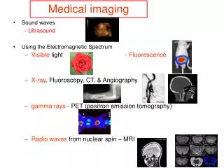
Medical Imaging. Mohammad Dawood Department of Computer Science University of Münster Germany. Medical Imaging Modalities. Introduction to Medical Imaging Modalities X-Ray CT MRT SPECT PET Ultrasound …. Electro-Magnetic Radiation. X-Ray CT MRT. X-Ray.


Medical Imaging
E N D
Presentation Transcript
Medical Imaging Mohammad Dawood Department of Computer Science University of Münster Germany
Introduction to Medical Imaging Modalities X-Ray CT MRT SPECT PET Ultrasound …
Production of X-Rays in Medical Diagnostic Cathode is heated Electrons are accelerated in an electric field Anode is used as target
X-Rays • produced when high velocity electrons collide with matter • Bremsstrahlung • Compton Scatter
X-Rays • 99% of the energy is converted into heat • -> targets with high melting points and atomic numbers • Rotating Anodes 3000-17000 rpm • Tungsten (Z=74, Tm=2757°, λ=1.3) • Rhenium (Z=75, Tm=2557°, λ=0.7)
X-Rays Attenuation effect
X-Rays • Attenuation effect • Wavelength • Atomic Number • Density • Thickness
X-Rays Contrast agents
X-Rays • Hounsfield scale (HU) • Scatter • Ray hardening
CT (Computed Tomography)
CT (Computed Tomography) Based on X-rays and attenuation effect
CT (Computed Tomography) 1st Generation CT Point by point 2nd Generation Detector array 3rd Generation Extended FOV 4th Generation Detector ring Electron Beam CT
MRT (Magnetic Resonance Tomography)
MRT (Magnetic Resonance Tomography Based on the spin of protons and an external magnetic field - Nuclei with odd number of protons have a spin - Due to electric charge they act as dipoles
MRT (Magnetic Resonance Tomography - dipoles are randomly distributed - When an external magnetic field (B0) is present the dipoles align themselves parallel or anti-parallel to it - more often parallel than anti-parallel 100,000:100,006 at 1.5 Tesla
MRT (Magnetic Resonance Tomography -the spins are not aligned exactly 0° or 180° to B0 - this leads to a precession - Larmor frequency
MRT (Magnetic Resonance Tomography) -net spin vector parallel to B0
MRT (Magnetic Resonance Tomography) - external RF signal (at larmor frequency) causes flip of M
MRT (Magnetic Resonance Tomography) appropriate external RF signal (at larmor frequency) causes flip of M into x-y plane precession induces a signal in a receiver coil T1 : time for re-alignment with B0 after the RF signal to 63%, usually in seconds
MRT (Magnetic Resonance Tomography • - T2 and T2* decay : time for decay to 37% in transverse magnetization, usually in milliseconds
MRT (Magnetic Resonance Tomography Selective Phase coding Frequency coding