Medical Imaging
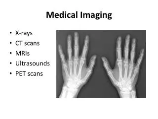
Medical Imaging. Tips for interpreting medical images. Objectives. Develop a repeatable pattern for interpretation Where to start / What to look for Recognizing normal Recognizing abnormal. Case 1. 2. 3. 1. Metal Mineral Soft tissue Fat Air. Dens Foramen transversarium


Medical Imaging
E N D
Presentation Transcript
Medical Imaging Tips for interpreting medical images
Objectives • Develop a repeatable pattern for interpretation • Where to start / What to look for • Recognizing normal • Recognizing abnormal
Case 1 2. 3. 1. Metal Mineral Soft tissue Fat Air • Dens • Foramen transversarium • Anterior arch of C1 • Lateral mass of C1 4. Recognizing normal: learn the names
4 considerations when faced with a medical image • Is the size of the structure normal? • Is the shape of the structure normal? • Is the position of the structure normal? • What is the density? (x-ray or CT) SIZE SHAPE POSITION DENSITY
1 Hard and Soft palate 2 X 3 4 5 • C5 vertebral body • Hyoid bone • Spinous process C4 • Dens • Occiput
Size Shape Position Density • What is wrong? • The vertebral body heights are abnormal • There is a fracture • There is malaligment • Nothing, this is normal
Grade 1 is 0–25% Grade 2 is 25–50% Grade 3 is 50–75% Grade 4 is 75–100% Over 100% is Spondyloptosis, when the vertabra completely falls off the supporting vertabra. Spondylolesthesis
Oblique lumbar x-ray Spondylolysis
Soft tissue swelling is an important sign in cervical trauma What are the normal measurements of the pre-cervical soft tissues at C2 and C6? “6 mm at 2, 22 mm at 6”
MRI Signal not density T1 – fluid is black T2 – fluid is white
Is there a problem with the size, shape, position or density of this structure?
Fracture description • Complete • Transverse • Mildly distracted (or separated) • Mid diaphysis • Left humerus • Not intra articular • Not open • Not angulated • Not comminuted
How would you describe these fractures?
MRI How would You describe this lesion? Benign versus malignant
B. A. Which of these images is normal?
Normal ACL Torn ACL anterior cruciate ligament
Test your knowledge The next cases have something wrong with size, shape, position and/or density Abnormal size Abnormal shape Abnormal position Abnormal density
Which bone is abnormal? How is it abnormal?
What is abnormal ? • Size • Shape • Position • Density
B. A. Which AP wrist x-ray is abnormal?
B. A. • What is abnormal here?? • Size • Shape • Position • Density
Do you see an abnormality here?
What to remember • You must learn the names of anatomic structures. • Be able to recite the 5 basic radiographic densities. • When looking at any structure, normal or abnormal, consider size, shape, position and density