Medical Imaging
Medical Imaging. Index. Medical Imaging Evolution of Medical Imaging Types of Medical Imaging Medical Radiography X-Ray Imaging Nuclear Imaging Bone Densitometry Magnetic Resonance Imaging (MRI) Ultrasound Neuroprosthetics Computed Tomography (CT) Fluoroscopy Echocardiography


Medical Imaging
E N D
Presentation Transcript
Index • Medical Imaging • Evolution of Medical Imaging • Types of Medical Imaging • Medical Radiography • X-Ray Imaging • Nuclear Imaging • Bone Densitometry • Magnetic Resonance Imaging (MRI) • Ultrasound • Neuroprosthetics • Computed Tomography (CT) • Fluoroscopy • Echocardiography • Digital Vascular Imaging • Positron Emission Tomography (PET) andRadionuclide Scanning
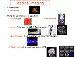
Medical Imaging Medical Imaging • Medical imaging is a discipline within the medical field which involves the use of technology to take images of the inside of the human body. These images are used in diagnostics, as teaching tools, and in routine healthcare for a variety of conditions. • Medical imaging is sometimes referred to as diagnostic imaging, because it is frequently used to help doctors arrive at a diagnosis, and there are a number of different types of technology used in medical imaging. The goal of medical imaging is to provide a picture of the inside of the body in a way which is as non-invasive as possible. An imaging study can be used to identify unusual things inside the body, such as broken bones, tumors, leaking blood vessels, and so forth. One of the most famous types of diagnostic imaging is the x-ray, which uses radiation to take a static image of a specific area of the body.
Evolution of Medical Imaging • Medical imaging began with the discovery of X-Rays by Wilhelm Conrad Röntgen in 1895 and his work showing that these rays could be used to peer inside the body and visualize bony structures. From this Nobel Prize winning discovery to the modern day, there are now many means by which the internal structures of the body can be assessed without the need for cutting it open.
Measurement and recording techniques which are not primarily designed to produce images, such as electroencephalography (EEG), magneto encephalography (MEG), electrocardiography (ECG) and others, but which produce data susceptible to be represented as maps (i.e. containing positional information), can be seen as forms of medical imaging.
Types of Medical Imaging • Medical Radiography • X-Ray Imaging • Nuclear Imaging • Bone Densitometry • Magnetic Resonance Imaging (MRI) • Ultrasound • Computed Tomography (CT) • Fluoroscopy • Echocardiography • Digital Vascular Imaging • Positron Emission Tomography (PET) andRadionuclide Scanning Types of Medical Imaging
Medical Radiography Medical Radiography • During a radiographic procedure, an x-ray beam is passed through the body. • A portion of the x-rays are absorbed or scattered by the internal structure and the remaining x-ray pattern is transmitted to a detector so that an image may be recorded for later evaluation. • The recoding of the pattern may occur on film or through electronic means Medical Radiography is considered a noninvasive procedure, which means that the process of acquiring a medical image does not penetrate the skin.
Radiography is used in many types of examinations and procedures where a record of a static image is desired. Some examples include • Dental examination • Verification of correct placement of surgical markers prior to invasive procedures • Mammography • Orthopedic evaluations • Spot film or recording during fluoroscopy • Chiropractic examinations
Digital Radiography MATRIX
X-Ray Imaging X-Ray Imaging • X-rays are waves that have a relatively high frequency along the electromagnetic spectrum. They are absorbed or transmitted by different body tissues in varying amounts, producing different shades of black and white on an x-ray image. The basic type of x-ray imaging is plain radiography. This involves an x-ray machine aimed at the patient's body with a recording plate positioned behind the region of interest. Once the machine delivers its radiation, the image is captured on the plate. This allows a physician to assess the bones for fractures, the abdomen for bowel obstruction, and the breasts for signs of cancer (mammography), among other applications. In general, bone appears white, soft tissue appears gray, and air appears black.
Nuclear Imaging Nuclear Imaging • Nuclear imaging is unique in that the means of visualization are not external waves delivered to the body. Rather, electromagnetic waves are emitted from within. In general, a radioactive marker is introduced into the patient's bloodstream and accumulates in the organ of interest. The marker gives off gamma rays that are detected by a special camera and processed into images by a computer. This type of imaging is also unique in that nuclear medicine physicians, not radiologists, are the ones who interpret nuclear imaging studies. Applications of nuclear imaging include perfusion of the heart, activity of the thyroid gland, and late-stage cancer.
Nuclear medicine images Lateral Reconstruction
Bone Densitometry • Bone density scanning is a form of X-ray technology used to measure a person’s bone loss.
Bone densitometry is most often used to diagnose osteoporosis, a condition that causes a gradual loss of calcium resulting in thinner and more fragile bones. Bone densitometry is also a preventative tool that tracks the effect of treatment for various conditions that cause bone loss.
Image Digitization Transducer Digitization with Matrix Arraying Scheme Windowing Inversion Image Sharpening
MRI Scanners Magnetic Resonance Imaging • MRI scanners, like X-rays and CT scanners, are basically machines doctors use to take pictures of your insides so that they can figure out what’s ailing you. • But MRI doesn’t involve ionizing radiation, as do X-rays and CT scans. Rather, MRI takes advantage of something you have plenty of in your body: water. It is far more flexible than X-rays and CT scans, and can generate three dimensional images in any orientation and at any depth in the body.
Residual Tumor Detection 1. Pre-opt MRI of tumor 2. Intraoperative MRI, residual tumor 3. Post-opt, no tumor
Ultrasound Imaging Ultra Sound Imaging • Ultrasound imaging uses high-frequency sound waves to produce detailed images of internal structures of the body. • Ultrasound imaging is used by physicians to get valuable information that will help them when it comes to diagnosing certain types of illnesses. • One such use is to determine the blood flow in a patients blood vessels. One drawback of ultrasound imaging is the fact that sound does not travel well through bone.
Ultrasound is produced and detected with a transducer, composed of one or more ceramic elements with electromechanical (piezoelectric) properties. • The ceramic element converts electrical energy into mechanical energy to produce ultrasound and mechanical energy into electrical energy for ultrasound detection.
Over the past several decades, the transducer assembly has evolved considerably in design, function, and capability, from a single-element resonance crystal to a broadband transducer array of hundreds of individual elements. • A simple single-element, plane-piston source transducer has major components including the • Piezoelectric material • Marching layer • Backing block • Acoustic absorber • Insulating cover • Sensor electrode • Transducer housing
Generic Ultrasonic Imaging System • Transmitter –Programmable apodization, delay control and frequency control. –Arbitrary receive direction. •Image processing: –Pre-detection filtering. –Post-detection filtering. •Full gain correction: analog and digital. •Scan converter: various scan format. –Arbitrary waveform. –Programmable transmit voltage. –Arbitrary firing sequence. –Programmable apodization, delay • Receiver
Image Data Acquisition c.f. Bushberg, et al. The Essential Physics of Medical Imaging, 2nd ed., p. 501.
Ultrasound • Heart and blood vessels, incl. the abdominal aorta and its major branches • Liver • Gallbladder • Spleen • Pancreas • Kidneys • Bladder • Eyes • Thyroid and parathyroid glands
Image Digitization - ADC Scanning Sampling Quantization
Ultrasound • Ultrasound waves do not pass through air; therefore an evaluation of the stomach, small intestine and large intestine may be limited. Intestinal gas may also prevent visualization of deeper structures such as the pancreas and aorta. • Patients who are obese are more difficult to image because tissue attenuates (weakens) the sound waves as they pass deeper into the body. Ultrasound has difficulty penetrating bone and therefore can only see the outer surface of bony structures and not what lies within.
Image Manipulation WINDOWING INVERSION IMAGE SHARPENING
Computed Tomography Computed Tomography Computed Tomography is a form of medical imaging using tomography that is created by computer processing. CT’s can be used to generate three-dimensional images of the inside of the human body. It does this by taking multiple two-dimensional X-ray images around a single axis. This data can then be manipulated to form an image of different body structures.
Computed Tomography (CT) is a powerful nondestructive evaluation (NDE) technique for producing 2-D and 3-D cross-sectional images of an object from flat X-ray images. Characteristics of the internal structure of an object such as dimensions, shape, internal defects, and density are readily available from CT images.
Unlike conventional radiography, in CT the patient lies on a couch that moves through into the imaging gantry housing the x-ray tube and an array of specially designed "detectors". • Depending upon the system the gantry rotates for either one revolution around the patient or continuously in order for the detector array to record the intensity of the remnant x-ray beam. • CT on the other hand reconstructs images in a variety of body planes the most usual being the axial or cross sectional plane. • Because of the large range of the CT number scale and the fact that the image is digital, it is possible to manipulate the display to show the underlying soft tissues with enhanced contrast as well as the bony structures. These recordings are then computer processed to produce images never before thought possible. Scanners today are capable of gathering even more data about the body structure in a time span that is measured in seconds thereby enhancing its clinical usefulness.
Why it is performed CT rapidly creates detailed pictures of the body, including the brain, chest, spine, and abdomen. • The test may be used to Diagnose an infection Guide a surgeon to the right area during a biopsy Identify masses and tumors, including cancer Study blood vessels
Risks • Being exposed to radiation • Allergic reaction to contrast dye • CT scans do expose you to more radiation than regular x-rays. Having many x-rays or CT scans over time may increase your risk for cancer
BASIC DATA AQUSITION SCHEME IN CT ANALOG ADC
Fluoroscopy Fluoroscopy • If a doctor needs real-time moving images of internal bodily structures, then they will use an imaging technique called fluoroscopy. • The process of fluoroscopy consists of using a radiation and a fluorescent screen. This form of imaging exposes the patient to extremely high forms of ionized radiation.
Echocardiography Echocardiography • An echocardiography is a medical imaging procedure that uses sound waves to create a moving picture of an organ, usually the heart. • Doctors use it because it gives a detailed picture and exposes the patient to zero radiation. • An echocardiogram enables the doctor to see the heart beating, rather than just looking at a picture of the heart. It also provides a detailed moving image of the heart valves that provide even more useful information to the doctor when attempting to diagnose an illness.
Types • Transthoracic Echocardiography • Stress Echocardiography • Trans-esophageal Echocardiography • Fetal Echocardiography • Three-Dimensional Echocardiography