Download
1 / 28
310 likes | 385 Vues
Discover the innovative world of modern medical imaging technologies, their applications in healthcare, and the collaborative efforts with various scientific fields. Learn about Magnetic Resonance Imaging (MRI), X-ray Imaging, Computed Tomography (CT), Positron Emission Tomography (PET), and Ultrasound. Through examples and detailed explanations, grasp the significance of spatial resolution and contrast in generating accurate medical images for diagnosis and treatment evaluation. Dive into the realm of medical imaging and its pivotal role in advancing healthcare practices.

E N D
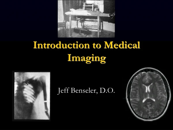
Introduction to Modern Medical Imaging Allen T. Newton, Ph.D. Institute of Imaging Science, Department of Radiology Vanderbilt University PAVE 2014
Medical Imaging • The goal is to better visualize structure or function in the living or non-living organism, animal or human • Involves collaborations with mathematicians, physicists, engineers, chemists and biologists • Work with physicians to meet their diagnostic and treatment evaluation needs • Look at a couple of key components of medical imaging • Look at some examples from MRI, ultrasound, CT
Chemistry Physics Imaging Science Biology Medicine Computer Science Math Engineering
Radiologists (MD) 4 yrs of college 4 yrs of medical school Physicist/engineer (PhD) 4 yrs of college 5-7 yrs of graduate school improve interpret Medical Imaging create Technologists (RT) 2-4 yrs of college 2 yrs of technical school
How do we make medical images? http://www.andor.com/image_lib/lores/introduction
The Major Imaging Modalities • Magnetic Resonance Imaging (MRI) • X-ray Imaging • Computed Tomography (CT) • Positron Emission Tomography (PET) • Ultrasound (US)
The Major Imaging Modalities • Magnetic Resonance Imaging (MRI) • X-ray Imaging • Computed Tomography (CT) • Positron Emission Tomography (PET) • Ultrasound (US)
The Major Imaging Modalities • Magnetic Resonance Imaging (MRI) • X-ray Imaging • Computed Tomography (CT) • Positron Emission Tomography (PET) • Ultrasound (US)
The Major Imaging Modalities • Magnetic Resonance Imaging (MRI) • X-ray Imaging • Computed Tomography (CT) • Positron Emission Tomography (PET) • Ultrasound (US)
So, you are playing soccer… • … you collide with another player, and collapse in pain on the ground • You have pretty intense pain in your lower left leg • How bad is it and how do we find out?
So, you go to the Emergency Room… • … and the attending physician orders an X-ray of your injured leg • What are they looking for? • What will the X-ray show? The X-ray shows a bad break in your tibia General Message: What is the problem? Can imaging solve the problem? How? Image adapted from http://www.gentili.net/image.asp
Spatial Resolution 32x32, 7.5 mm2 64x64, 3.75 mm2 128x128, 1.87 mm2 256x256, 0.93 mm2
Spatial resolution 7T MRI, 0.6x0.6x1.0 mm3 resolution
Magnetic Resonance imaging Coronal Sagittal • Can generate 2D and 3D views in any plane • Uses no ionizing radiation • Extremely versatile modality • Spatial resolution: humans~100, animals~25 microns Axial
Magnetic Resonance imaging MRI showing blood vessels in brain
Bright in coherent white matter Darker where 1) Fibers diverge/cross, or 2) No preferred orientation Magnetic Resonance imaging Assessing brain white matter tracks • Orientation is color code by direction • Red = Right/Left • Green = Anterior/Posterior • Blue = Superior/Inferior • Reveals structure within white matter Adam Anderson, Ph.D.
Magnetic Resonance imaging Finding white matter tracks Establishes connections between different brain regions Adam Anderson, Ph.D.
MRI in cancer imaging Signal Intensity time courses 3D rendering of tumor anatomical MRI
X-rays in cancer imaging, 1/2 Mammogram displaying calcification, increased density Mrs. Röntgen's hand, the first X-ray picture of the human body ever taken
X-rays in cancer imaging, 2/2 Munden, et al, Radiology, 2005; 237:803-18 • Standard radiography yields a 2-D projection of a 3D object, whereas CT allows for true 3-D image acquisition • CT acquires a series of projections from many angles around the subject; each set of projections is then reconstructed via a “backprojection” algorithm • Developed by Sir Godfrey Houndsfield, won 1972 Nobel Prize in Medicine/Physiology • Spatial resolution: humans~200 microns, animals~35 micron www.radiologyinfo.org/
PET in cancer imaging, 1/2 • Some radioactive isotopes (18F, 15O, etc) emit positrons: • Such elements can be incorporated into metabolically relevant compounds • Emitted positron encounters electron, they annihilate leaving 2 photons traveling in opposite directions which are measured by ring of detectors • Most common PET tracer is FDG (flourodeoxyglucose), a glucose analog 18FDG (blood) 18FDG (tissue) 18FDG-6-PO4 (cells) X • Images generated using very similar computations as in CT • Spatial resolution: humans and animals ~2 mm
PET in cancer imaging, 2/2 CTPETCT/PET Dominique Delbeke www.breastcancer.org
This week Today: Overview Tuesday: X-Ray & Computed Tomography (CT) Wednesday: Ultrasound, SPECT, PET Thursday: MRI Friday: fMRI lab !!!!!!