Download

1 / 13

E N D
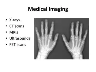
X-Rays • What is a Routine X-Ray? • The X-ray has been called one of the most significant advances in all of medical history. It is used in many different ways in medical diagnosis. An x-ray image is produced when a small amount of radiation passes through the body and strikes a sheet of sensitive film placed on the other side of the body. The ability of x-rays to penetrate tissues and bones varies according to the tissue's composition and mass. Bone, which contains calcium, does not let much radiation through and results in white images on the x-ray film. The lungs, which are filled with air, allow nearly all x-rays to strike the film resulting in a black film image.
Medical Imaging Techniques • X-rays
CT Scan • What is a CT scanner?A CT (computerised tomography) scanner is a special kind of X-ray machine. Instead of sending out a single X-ray through your body as with ordinary X-rays, several beams are sent simultaneously from different angles. How does a CT scanner work?The X-rays from the beams are detected after they have passed through the body and their strength is measured.Beams that have passed through less dense tissue such as the lungs will be stronger, whereas beams that have passed through denser tissue such as bone will be weaker. A computer can use this information to work out the relative density of the tissues examined. Each set of measurements made by the scanner is, in effect, a cross-section through the body
What is a PET scan? • A positron emission tomography (PET) scan is a unique type of imaging test that helps doctors see how the organs and tissues inside your body are actually functioning. • The test involves injecting a very small dose of a radioactive chemical, called a radiotracer, into the vein of your arm. The tracer travels through the body and is absorbed by the organs and tissues being studied. Next, you will be asked to lie down on a flat examination table that is moved into the center of a PET scanner—a doughnut-like shaped machine. This machine detects and records the energy given off by the tracer substance and, with the aid of a computer, this energy is converted into three-dimensional pictures. A physician can then look at cross-sectional images of the body organ from any angle in order to detect any functional problems. • What problems can a PET scan detect?A PET scan can measure such vital functions as blood flow, oxygen use, and glucose metabolism, which helps doctors identify abnormal from normal functioning organs and tissues. The scan can also be used to evaluate the effectiveness of a patient’s treatment plan, allowing the course of care to be adjusted if necessary. • Currently, PET scans are most commonly used to detect cancer, heart problems (such as coronary artery disease and damage to the heart following a heart attack), brain disorders (including brain tumors, memory disorders, seizures) and other central nervous system disorders. • How is a PET scan different from a CT or MRI scan?One of the main differences between PET scans and other imaging tests like CT scan or magnetic resonance imaging (MRI) is that the PET scan reveals the cellular level metabolic changes occurring in an organ or tissue. This is important and unique because disease processes often begin with functional changes at the cellular level. A PET scan can often detect these very early changes whereas a CT or MRI detect changes a little later—as the disease begins to cause changes in the structure of organs or tissues
What is an MRI scan? • MRI (magnetic resonance imaging) is a fairly new technique that has been used since the beginning of the 1980s.The MRI scan uses magnetic and radio waves, meaning that there is no exposure to X-rays or any other damaging forms of radiation. How does an MRI scanner work? The patient lies inside a large, cylinder-shaped magnet. Radio waves 10,000 to 30,000 times stronger than the magnetic field of the earth are then sent through the body. This affects the body's atoms, forcing the nuclei into a different position. As they move back into place they send out radio waves of their own. The scanner picks up these signals and a computer turns them into a picture. These pictures are based on the location and strength of the incoming signals. Our body consists mainly of water, and water contains hydrogen atoms. For this reason, the nucleus of the hydrogen atom is often used to create an MRI scan in the manner described above. What does an MRI scan show? Using an MRI scanner, it is possible to make pictures of almost all the tissue in the body. The tissue that has the least hydrogen atoms (such as bones) turns out dark, while the tissue that has many hydrogen atoms (such as fatty tissue) looks much brighter. By changing the timing of the radiowave pulses it is possible to gain information about the different types of tissues that are present. An MRI scan is also able to provide clear pictures of parts of the body that are surrounded by bone tissue, so the technique is useful when examining the brain and spinal cord. Because the MRI scan gives very detailed pictures it is the best technique when it comes to finding tumours (benign or malignant abnormal growths) in the brain. If a tumour is present the scan can also be used to find out if it has spread into nearby brain tissue. The technique also allows us to focus on other details in the brain. For example, it makes it possible to see the strands of abnormal tissue that occur if someone has multiple sclerosis and it is possible to see changes occurring when there is bleeding in the brain, or find out if the brain tissue has suffered lack of oxygen after a stroke. The MRI scan is also able to show both the heart and the large blood vessels in the surrounding tissue. This makes it possible to detect heart defects that have been building up since birth, as well as changes in the thickness of the muscles around the heart following a heart attack. The method can also be used to examine the joints, spine and sometimes the soft parts of your body such as the liver, kidneys and spleen.
What is General Ultrasound Imaging? • Ultrasound imaging, also called ultrasound scanning or sonography, is a method of obtaining images from inside the human body through the use of high-frequency sound waves. The reflected sound wave echoes are recorded and displayed as a real-time visual image. No ionizing radiation (x-ray) is involved in ultrasound imaging. Obstetric ultrasound refers to the specialized use of sound waves to visualize and thus determine the condition of a pregnant woman and her embryo or fetus. • Ultrasound is a useful way of examining many of the body's internal organs, including but not limited to the heart, liver, gallbladder, spleen, pancreas, kidneys and bladder. Because ultrasound images are captured in real time, they can show movement of internal tissues and organs and enable physicians to see blood flow and heart valve functions. This can help to diagnose a variety of heart conditions and to assess damage after a heart attack or other illness.