Download
1 / 13
140 likes | 373 Vues
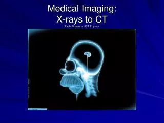
Medical Imaging: X-rays to CT Zach Simmons UST Physics. History. X-Rays: Wilhelm Conrad R öntgen, 1895 Medical applications immediately apparent Safety concerns not immediately apparent Basically remained unchanged until CT

E N D
History • X-Rays: Wilhelm Conrad Röntgen, 1895 • Medical applications immediately apparent • Safety concerns not immediately apparent • Basically remained unchanged until CT • Computerized Tomography (CT) : first practical realization appeared in the 1970’s • First commercial CT scanner: invented in 1971 by Godfrey Hounsfield, thanks to EMI and the Beatles
X-Rays:Bremsstrahlung cathode anode
OK, so: • Different types of tissue attenuate x-rays at different rates, causing there to be contrast in intensity transmitted to the resulting image depending on the tissue • Beer’s law: • Some example attenuation coefficients: µair =0, µbone =.48/cm, µmuscle =.180/cm -to give you an idea: about 40% attenuation in 1cm of bone
X-Ray Radiography • Projection of a 3D person onto a 2D film
CT (Computerized Tomography) is where it gets interesting: -Yields slices, instead of a “shadow” -This is the simplest scanning configuration; there’s lots more complicated geometries and this isn’t how modern CT scanners work.
Improvements/other: • 1980’s Spiral CT and different geometries • Fluoroscopy • Higher resolution • Faster scans also improves quality • 3-D image reconstruction
Sources: Books: Cho, Z.H. Foundations of Medical Imaging, New York: John Wiley & Sons, Inc. 1993. Webb, S. Ed. The physics of Medical Imaging, New York: Adam Hilger, 1988. Pics: Slide 1: http://www.lgmsworld.de/de/downloads/hintergrundbilder/homer/homer_1024.jpg Slide 3: http://hyperphysics.phy-astr.gsu.edu/hbase/quantum/xtube.html Slide 5: http://www.dmacgroup.com/Chest%20X-ray.jpg http://www.le.ac.uk/pathology/teach/CA/Cases/examination8f1.htm Slide 6: http://www.e-radiography.net/ibase5/5_Equipment/ Slide 7: Webb, p.100 http://medinfo.ufl.edu/cme/geri/images/ct5.jpg Slide 8: Webb, p.107 Slide 12: http://147.163.65.155/mambo/index.php?option=content&task=view&id=42