Patient-Specific 3D Biomechanical Modeling for Obstructive Sleep Apnea Diagnosis
This document outlines the OPAL Project, which focuses on creating patient-specific 3D biomechanical models for individuals with Obstructive Sleep Apnea (OSA). The project employs a workflow involving imaging, image processing, reference model generation, and biomechanical modeling to enhance diagnosis and treatment. Utilizing high-resolution data from various sources, the aim is to develop accurate representations of anatomical structures such as the airway, tongue, and palate. These models facilitate simulations that are crucial for clinicians in understanding and addressing OSA.

Patient-Specific 3D Biomechanical Modeling for Obstructive Sleep Apnea Diagnosis
E N D
Presentation Transcript
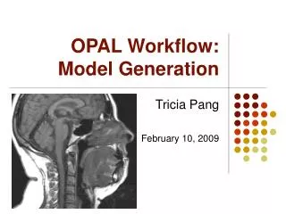
OPAL Workflow: Model Generation Tricia Pang February 10, 2009
Motivation • ArtiSynth [1]:3D Biomechanical Modeling Toolkit • Ideally: • Model derived from single subject source • High resolution model
Motivation • Obstructed sleep apnea (OSA) disorder • Caused by collapse of soft tissue walls in airway • Ideally: • Ability to run patient-specific simulations to help diagnosis • Quick and accurate method of generating model Credit: Wikipedia
OPAL Project • Dynamic Modeling of theOral, Pharyngeal and Laryngeal (OPAL)Complex for Biomedical Engineering • Patient-specific modeling and model simulation for study of OSA • Tools for clinician use in segmenting image and importing to ArtiSynth • Come up with protocol, tools/techniques and modifications needed for end-to-end process
OPAL Project 3D Medical Data Biomechanical Model
Workflow Stages 1. Imaging 2. Image processing & reconstruction 3. Reference model generation 4. Patient-specific model fitting 5. Biomechanical model
Workflow Stages 1. Imaging 2. Image processing & reconstruction 3. Reference model generation 4. Patient-specific model fitting 5. Biomechanical model
Stage 1: Imaging • Structures • Tongue • Soft palate • Hard palate • Epiglottis • Pharyngeal wall • Airway • Jaw • Teeth
Data Source Dental Appliancew/ Markers Cone CT of Dental Cast MRI Credit: Klearway, Inc. Other:laser scans, planar/full CT scans, tagged MRI, ultrasound, fluoroscopy, cadaver data…
MRI & Protocol • Normal subject vs. OSA patients • Control vs. treatment (appliance)
Workflow Stages 1. Imaging 2. Image processing & reconstruction 3. Reference model generation 4. Patient-specific model fitting 5. Biomechanical model
Stage 2: Image processing & Reconstruction • N3 correction [2] (Non-parametric non-uniform intensity normalization) • Cropping • Cubic interpolation • Image registration & reconstruction (Bruno’s work) • Combining 3 data sets → high-quality data set
Workflow Stages 1. Imaging 2. Image processing & reconstruction 3. Reference model generation 4. Patient-specific model fitting 5. Biomechanical model
Stage 3:Reference Model Generation • Goal: High quality model • Focus on bottom-up semi-automatic segmentation approaches • eg. Livewire [3]
3D Livewire Seed points (forming contours) drawn in 2 orthogonal slice directions, and seed points automatically generated in third slice direction
Livewire ModelRefinement (Claudine & Tanaya) • Morphological operations • Contour smoothening(active contours [4]) • 3D surface reconstruction(non-parallel curve networks [5])
Workflow Stages 1. Imaging 2. Image processing & reconstruction 3. Reference model generation 4. Patient-specific model fitting 5. Biomechanical model
Stage 4:Patient-Specific Model Generation • Goal: Accurate model, generated with minimal user interaction • Focus on top-down or automated approaches • Morphological warping operations • Deformable model crawlers
Thin-Plate Spline Warping • Thin-plate spline (TPS) deformation [6]: interpolating surfaces over a set of landmarks based on linear and affine-free local deformation Reference Model Warp Result Warp field
TPS Warping, Phase 1 • User selects a point on both patient MRI and reference model • Hard to pinpoint landmarks on 3D model Patient MRI List of corresponding points Reference Model
TPS Warping, Phase 2 Reference MRI (has a pre-built 3D model) • Predefined landmarks shown on reference MRI, user selects equivalent point on patient MRI • Can be improved by automated point-matching Patient MRI
Chan-Vese Active Contours • Highly automated method • Combine 2D segmentation of axial slices in Matlab • User-indicated start point • Iterate sequentially using previous segmentation as starting contour for Chan-Vese active contours [7] Automated AC on axial(2 minutes) Livewire 3D (~2 hours) Livewire +post processing
Deformable Organism Crawler • Automatically segment airway by growing a tubular organism, guided by image data and a priori anatomical knowledge • Developed in I-DO toolkit [8] • Advantages: • Analysis and labeling capabilities • Ability to incorporate shape-basedprior knowledge • Modular hierarchical development framework
Workflow Stages 1. Imaging 2. Image processing & reconstruction 3. Reference model generation 4. Patient-specific model fitting 5. Biomechanical model
Stage 5:Biomechanical Model • Import surface mesh into ArtiSynth • Work in progress • Challenges: • Determining “rest” position from inverse modeling • Defining interior nodes and muscle end points
Challenges inSegmentation • Medical image data quality • Bottom-up methods: Need for general procedure and abstraction from anatomy being segmented • Top-down methods: Need good atlas model • Validation with gold standard segmentation
Future Directions in Segmentation • Deformable organism crawler • Automated morphing of reference model into patient model • Additions to Livewire • Oblique slices • Sub-pixel resolution • Convert to graphics implementation • Add smoothness by regularization(eg. by spline, a priori model, …)
References [1] Fels, S., Vogt, F., van den Doel, K., Lloyd, J., Stavness, I., and Vatikiotis-Bateson, E. Developing Physically-Based, Dynamic Vocal Tract Models using ArtiSynth. Proc. Int. Seminar Speech Production (2006), 419-426. [2] Sled, G., Zijdenbos, A. P., and Evans, A. C. Non-parametric method for automatic correction of intensity nonuniformity in MRI data. IEEE Trans. in Medical Imaging17, 1 (1998), 87-97. [3] Poon, M., Hamarneh, G., and Abugharbieh, R. Effcient interactive 3d livewire segmentation of complex objects with arbitrary topology. Comput. Med Imaging and Graphics (2009), in press. [4] Hamarneh, G., Chodorowski, A., and Gustavsson, T. Active Contour Models: Application to Oral Lesion Detection in Color Images. IEEE International Conference on Systems, Man, and Cybernetics 4 (2000), 2458 -2463. [5] Liu, L., Bajaj, C., Deasy, J. O., Low, D. A., and Ju, T. Surface reconstruction from non-parallel curve networks. Eurographics 27, 2 (2008), 155-163. [6] Bookstein, F. L. Principal Warps: Thin-Plate Splines and the Decomposition of Deformations. IEEE Transactions on Pattern Analysis and Machine Intelligence 11, 6 (1989), 567-585. [7] Chan, T., and Vese, L. Active contours without edges. IEEE Transactions on Image Processing 10, 2 (2001), 266-277. [8] McIntosh, C. and Hamarneh, G. I-DO: A “Deformable Organisms” framework for ITK. Medical Image Analysis Lab, SFU. Release 0.50.