Electronic Supplementary Information for:
Electronic Supplementary Information for: Delivery, stabilization, and spatiotemporal activation of cargo molecules in cells with positively charged nanoparticles Shuhei Murayama, Taihei Nishiyama, Kaihei Takagi, Fumi Ishizuka, Tomofumi Santa and Masaru Kato *

Electronic Supplementary Information for:
E N D
Presentation Transcript
Electronic Supplementary Information for: Delivery, stabilization, and spatiotemporal activation of cargo molecules in cells with positively charged nanoparticles Shuhei Murayama,Taihei Nishiyama,Kaihei Takagi, Fumi Ishizuka, Tomofumi Santa and Masaru Kato * Graduate School of Pharmaceutical Sciences, Global COE Program (CMSI) and Leading program (GPLLI), The University of Tokyo, 7-3-1 Hongo, Bunkyo-ku, Tokyo 113-0033, Japan * To whom corresponding should be addressed: E-mail: masaru-kato@umin.ac.jp
Methods Materials. Tetra-poly(ethyl glycol)-amine (SUNBRIGHT PTE-050PA; Mn, 5328 g/mol) was purchased from NOF Corporation (Tokyo, Japan). N,N,N',N'-Tetramethylethylenediamine (TEMED), dichloromethane (DCM), acryloyl chloride (AC), triethylamine (TEA), ammonium persulfate (APS), tris(hydroxymethyl) aminomethane, hydrochloric acid, methanol, acetone, 1- butyl alcohols, diethyl ether, 4-(4,6-dimethoxy-1,3,5-triazin-2-yl)-4-methylmorpholinium chloride n-hydrate (DMT-MM), sodium bicarbonate, trypsin (porcine), rhodamine B, and chlorpromazine hydrochloride were purchased from Wako Pure Chemical Industries (Osaka, Japan). Methyl-b-cyclodextrin was purchased from Tokyo Chemical Industry (Tokyo, Japan). 4-[4-(1-Hydroxyethyl)-2-methoxy-5-nitrophenyl] butyric acid, polypropylenimine tetramine dendrimer, generation 1.0 (DAB-Am-4), fluorescein sodium salt, amiloride hydrochloride hydrate and fluorescein isothiocyanate conjugate with bovine albumin were purchased from Sigma-Aldrich (St. Louis, MO). Water was purified with a Milli-Q apparatus (Millipore, Bedford, MA). Boron dipyrromethene (BODIPY)-casein was purchased from invitrogen (California, U.S.A). Synthesis of photocleavable linker (PEG-photo-Ac) The photocleavable linker, PEG-photo-Ac, was prepared based on the procedure of similar molecules.S1 Photo-Ac (5 mmol) was dissolved in methanol and stirred. Then, tetra poly(ethylene glycol)-amine (tetra-PEG-amine; 1 mmol) was added and stirred until all reactants were dissolved. After the addition of DMT-MM, the stir was stopped.4, The reaction was done at room temperature for overnight. The product was precipitated in diethyl ether on ice and filtered. The collected substance was washed with diethyl ether and dissolved in water. The aqueous solution was dialyzed (SpectraPor6, CO 1000 gmol-1) and freeze-dried to yield the tetra-acrylated PEG referred to as PEG-photo-Ac (Scheme 1). The 1H NMR spectrum of the PEG-photo-Ac prepared in this procedure was the same as that was prepared by our previous study. 3
Preparation of the nanoparticles containing encapsulated molecules DAB-Ac was prepared as described in our previous report.5 We prepared solvents of PEG-photo-Ac and DAB-Ac at various proportions of DAB-Ac (0, 1, 5, 10, 20, and 50%). To 100 mL of each solution, we added 100 mL of a solvent of the molecule to be encapsulated (fluorescein, rhodamine B, BODIPY-casein, or fluorescein-albumin), 0.1 M APS (50 mL), and 0.1 M TEMED in 1 M Tris/HCl buffer (pH 7.0, 50 mL) in that order. The solutions were then stirred for 20 min at room temperature. Diameter and zeta potential measurements by DLS Delsa Nano (Beckman Coulter. Inc. Brea CA, USA) was used for the measurement of diameters and zeta potentials of the prepared nanoparticles. The prepared nanoparticle solution was filtrated by filter (Ultrafree-MC centrifugal filter) and diluted by water (x 10) before the measurement. Cell culture The human hepatocyte carcinoma cells (HuH-7), Cos-7 cells and normal human umbilical vein endothelial cells (HUVEC) from Cambrex Corporation (East Rutherford, NJ) were cultured in a humidified atmosphere of 95% air and 5% CO2 at 37°C in Dulbecco`s Modified Eagle`s Medium (DMEM, Sigma-Aldlich) supplemented with 1% (v/v) penicillin-streptomycin solution (x 100) (Wako) and 10% (v/v) fetal bovine serum (FBS, invitorogen). A solution of 10 ng/mL b-FGF (Sigma-Aldrich) was added to the medium for HUVEC. For each experiment, cells at 80-90% confluence were harvested by trypsin/EDTA (Wako) digestion, washed and re-suspended in fresh growth medium with FBS at a cell concentration of 106 viable cells/culture dish (75 cm2). HuH-7 and Cos-7 were cultured in 35 mm glass bottom dish. HUVEC was cultured in 35 mm glass dish coated with collagen. Internalization of the nanoparticles to the cell The cultured dish was washed by DMEM without serum and added another DMEM (2 mL) to the dish. Before the addition to the cultured dishes, the prepared nanoparticles were filtered through an Ultrafree-MC centrifugal filter (0.22 mm GV Durapore; Millipore, Billerica MA, USA). Then 50 mL of filtrate was dropped to the dish. Then stand the dish for 20 min for internalization. After that, the sample was washed 3 times by the medium.
UV irradiation and microscopy observation The cells were irradiated with UV light (Aicure UV20, Panasonic, Osaka, Japan) for 20 s (0.25 W/cm2), and then confocal laser scanning microscopy (CLSM) images of the cells were obtained with an LSM 510 microscope (Carl Zeiss AG., Oberkochen, Germany). Inhibitor assay HuH-7 cells in DMEM with serum were seeded in a dish one day before the assay. After overnight incubation, the dish was washed by DMEM without serum and added 2 mL of DMEM. We added 500 mL of endcytosis inhibitor (chlorpromazine (6 mg/mL) or methyl-b-cyclodextrin (13 mg/mL) or amiloride (0.8 mg/mL)) to the culture dishesS2 and then 50 mL of nanoparticles solution was dropped and allowed to stand for 20 min at room temperature. After the cells were washed 3 times by the medium, the cells were observed by an LSM 510 microscope. Analysis of florescence images Internalization of the nanoparticles in HuH-7 cells was evaluated by means of confocal laser scanning microscopy. The fluorescence intensity in the images was analyzed with ImageJ software (ver. 1.45s; http://rsbweb.nih.gov/ij/index.html). The mean gray value of each cell was calculated from six images. The mean gray values of rhodamine B and fluorescein-albumin were calculated from fluorescence image of HuH-7 cells before irradiation, because free rhodamine B was unstable within cell. The mean gray value of BODIPY-casein was calculated from image of HuH-7 that obtained 2 hours later from irradiation, because we need to wait for the digestion reaction of BODIPY-casein by endogenous protease. References S1 A. M. Kloxin, A. M. Kaskko, C. N. Salinas and K. S. Anseth, Science 2009, 324, 59-63. S2 P.G. Cammisotto, M. Bendayan, A. Sané, M. Dominguez and C. Garofalo, É. Levy, Int. J. Cell Bio.2010, 2010, 928169 (13 pages).
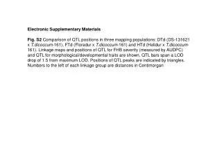
Fig. S1 Fluorescence image of HUVECtreated with nanoparticles containing fluorescein. Scale bars = 20 mm. Fig. S2 Fluorescence image of HuH-7 cell treated with nanoparticles containing fluorescein-albumin. Scale bars = 20 mm.
60 40 Mean Gray Value 20 0 1 day 2 day 3 day 4 day before UV after UV before UV after UV before UV after UV before UV after UV Fig. S3 Change of relative mean gray values of cells treated with nanoparticles containing BODIPY-casein before and after irradiation.
1day 2day 3day 4day Fig. S4 Merged images of HuH-7 cell treated with nanoparticles. Scale bars = 20 mm.